纺织学报 ›› 2024, Vol. 45 ›› Issue (09): 1-9.doi: 10.13475/j.fzxb.20230701601
• 纤维材料 • 下一篇
房磊1, 刘秀明1, 贾娇娇2, 蔺志浩3, 任燕飞2, 侯凯文4, 巩继贤1, 扈延龄3( )
)
FANG Lei1, LIU Xiuming1, JIA Jiaojiao2, LIN Zhihao3, REN Yanfei2, HOU Kaiwen4, GONG Jixian1, HU Yanling3()
摘要:
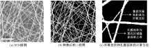
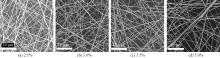
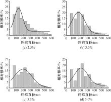
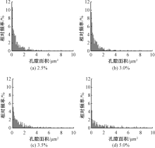
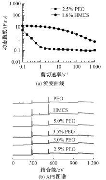
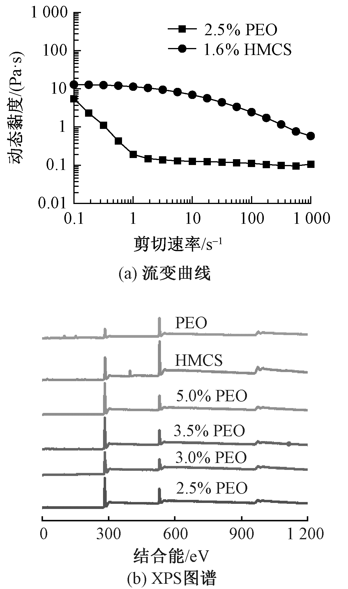
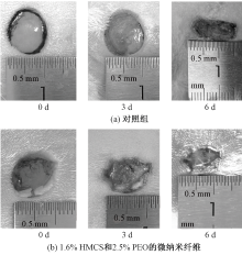
高分子量壳聚糖因具有良好的抗菌性、可促进细胞和组织生长以及可降解等特性,被认为是促进伤口愈合的优良材料;然而高分子量壳聚糖纺丝液黏度大,用常规静电纺丝法难以制成微纳米纤维。为解决这个问题,采用溶液喷射纺丝法制备了高分子量壳聚糖/聚氧化乙烯微纳米纤维,纺丝液含有质量分数为1.6%的高分子量壳聚糖和2.5%~5.0%的聚氧化乙烯(相对分子质量为10万)。所制备的微纳米纤维呈直线形,表面呈不光滑波纹状。通过MatLab对扫描电子显微镜照片进行识别和计算,结果表明,当聚氧化乙烯质量分数从2.5% 增加到5.0%时,纤维平均直径从133 nm增加到210 nm,直径分布变宽;微纳米纤维材料中大孔隙增多,孔隙率在0.69左右,变化不大;X射线光电子能谱和透射电子显微镜分析结果表明,所制备的高分子量壳聚糖/聚氧化乙烯喷射纺丝微纳米纤维具有皮芯结构,其中,聚氧化乙烯位于皮层,高分子量壳聚糖构成微纳米纤维的内芯;动物实验初步证明了高分子量壳聚糖皮芯结构微纳米纤维可以促进伤口愈合。
中图分类号:
| [1] | OMAR Belal A, ELMASRY Ragab, EITA Ahmed, et al. Upgrading the preparation of high-quality chitosan from Procambarus clarkii wastes over the traditional isolation of shimp chitosan[J]. Saudi Journal of Biological Science, 2022, 29(2): 911-919. |
| [2] | WEIßPFLOG Janek, VEHLOW David, MÜLLER Martin, et al. Characterization of chitosan with different degree of deacetylation and equal viscosity in dissolved and solid state: insights by various complimentary methods[J]. International Journal of Biological Macromolecules, 2021, 171: 242-261. |
| [3] | YU Ling, DOU Shubin, MA Jinghan, et al. An antimicrobial peptide-loaded chitosan/polyethylene oxide nanofibrous membrane fabricated by electrospinning technology[J]. Frontiers in Materials, 2021. DOI: 10.3389/fmats.2021.650223. |
| [4] | LI Jianhui, FU Jimin, TIAN Xiao, et al. Characteristics of chitosan fiber and their effects towards improvement of antibacterial activity[J]. Carbohydrate Polymers, 2022. DOI: 10.1016/j.carbpol.2021.119031. |
| [5] | MATICA, AACHMANN, TONDERVIK, et al. Chitosan as a wound dressing starting material: antimicrobial properties and mode of action[J]. International Journal of Molecular Sciences, 2019. DOI: 10.3390/ijms20235889. |
| [6] |
DI SANTO Mariana Carolina, ALAIMO Agustina, ACEBEDO Sofia Lorena, et al. Biological responses induced by high molecular weight chitosan administrated jointly with platelet-derived growth factors in different mammalian cell lines[J]. International Journal of Biological Macromolecules, 2020, 158: 953-967.
doi: S0141-8130(20)33177-9 pmid: 32423872 |
| [7] |
ALSARRA Ibrahim A. Chitosan topical gel formulation in the management of burn wounds[J]. International Journal of Biological Macromolecules, 2009, 45(1): 16-21.
doi: 10.1016/j.ijbiomac.2009.03.010 pmid: 19447254 |
| [8] | JONES Mitchell, KUJUNDZIC Marina, JOHN Sabu, et al. Crab vs. mushoom: a review of crustacean and fungal chitin in wound treatment[J]. Marine Drugs, 2020. DOI: 10.3390/md18010064. |
| [9] | HIGASHI Shougo, HIRAI Takayuki, MATSUBARA Masato, et al. Dynamic viscosity recovery of electrospinning solution for stabilizing elongated ultrafine polymer nanofiber by TEMPO-CNF[J]. Scientific Reports, 2020. DOI: 10.1038/s41598-020-69136-2. |
| [10] | TIWARI Sandeep Kumar, VENKATRAMAN Subbu S. Importance of viscosity parameters in electrospinning: of monolithic and core-shell fibers[J]. Materials Science and Engineering: C, 2012, 32(5): 1037-1042. |
| [11] | GREYLING Corinne Jean. Electrospinning of polyacrylonitrile nanofibres with additives: study of orientation and crystallinity[D]. Stellenbosch: University of Stellenbosch, 2010: 229. |
| [12] | AMARIEI N, MANEA L R, BERTEA A P, et al. The Influence of polymer solution on the properties of electrospun 3D nanostructures[J]. IOP Conference Series (Materials Science and Engineering), 2017. DOI: 10.1088/1757-899X/209/1/012092. |
| [13] | MA Lulu, DENG Li, CHEN Jianming. Applications of poly(ethylene oxide) in controlled release tablet systems: a review[J]. Drug Development and Industrial Pharmacy, 2013. DOI: 10.3109/03639045.2013.831438. |
| [14] | LEE Jin Ho, LEE Hai Bang, ANDRADE Joseph D. Blood compatibility of polyethylene oxide surfaces[J]. Progress in Polymer Science, 1995, 20: 1043-1079. |
| [15] | MANDY Stephen H. A new primary wound dressing made of polyethylene oxide gel[J]. Journal of Dermatologic Surgery and Oncology, 1983. DOI: 10.1111/j.1524-4725.1983.tb00778.x. |
| [16] | SALASSA John R, PEARSON Bruce W. Polyethylene oxide gel: a new intranasal dressing after septorhinoplasty[J]. Archives of Otorhinolaryngology-Head & Neck Surgery, 1991, 117:1365-1367. |
| [17] |
MA Lulu, DENG Li, CHEN Jianming. Applications of poly(ethylene oxide) in controlled release tablet systems: a review[J]. Drug Development and Industrial Pharmacy, 2014, 40(7):845-851.
doi: 10.3109/03639045.2013.831438 pmid: 24001212 |
| [18] | ZHANG Jianfeng, YANG Dongzhi, XU Fei, et al. Electrospun core-shell structure nanofibers from homogeneous solution of poly(ethylene oxide)/chitosan[J]. Macromolecules, 2009, 42(14): 5278-5284. |
| [19] |
PAKRAVAN Mehdi, HEUZEY Marie Claude, AJJI Abdellah. Core-shell structured PEO-chitosan nanofibers by coaxial electrospinning[J]. Biomacromolecules, 2012, 13(2): 412-421.
doi: 10.1021/bm201444v pmid: 22229633 |
| [20] | ASHOK K, BABU M, SANKAR A, et al. Nanofiber based dressing material mechanism in wound healing property[J]. International Journal of Zoological Investigations, 2021, 7(2): 949-954. |
| [21] | WANG Fadong, HU Shui, JIA Qingxiu, et al. Advances in electrospinning of natural biomaterials for wound dressing[J]. Journal of Nanomaterials, 2020. DOI: 10.1155/2020/8719859. |
| [22] | LIU Yukang, LI Chaofei, FENG Zhangbin, et al. Advances in the preparation of nanofiber dressings by electrospinning for promoting diabetic wound healing[J]. Biomolecules, 2022. DOI: 10.3390/biom12121727. |
| [23] | MEDEIROS Eliton S, GLENN Gregory M, KLAMCZYNSKI Artur P, et al. Solution blow spinning: a new method to produce micro- and nanofibers from polymer solutions[J]. Journal of Applied Polymer Science, 2009, 113(4): 2322-2330. |
| [24] | DADOL Glebert C, KILIC Ali, TIJING Leonard D, et al. Solution blow spinning (SBS) and SBS-spun nanofibers: materials, methods, and applications[J]. Materials Today Communications, 2020. DOI: 10.1016/j.mtcomm.2020.101656. |
| [25] |
HUANG Ya, BAI Xiaopeng, ZHOU Ming, et al. Large-Scale spinning of silver nanofibers as flexible and reliable conductors[J]. Nano Letters, 2016, 16(9): 5846-5851.
doi: 10.1021/acs.nanolett.6b02654 pmid: 27548808 |
| [26] |
MURPHY Ryan, TURCOTT Ashley, BANUELOS Leo, et al. SIMPoly: a MatLab-based image analysis tool to measure electrospun polymer scaffold fiber diameter[J]. Tissue Engineering Part C: Methods, 2020, 26(12): 628-636.
doi: 10.1089/ten.TEC.2020.0304 pmid: 33256558 |
| [27] | WANG Yan, YOKOTA Tomoyuki, SOMEYA Takao. Electrospun nanofiber-based soft electronics[J]. NPG Asia Materials, 2021. DOI: 10.1038/s41427-020-00267-8. |
| [28] | RYU Hyun Il, KOO Min Seok, KIM Seokjun, et al. Uniform-thickness electrospun nanofiber mat production system based on real-time thickness measurement[J]. Scientific Reports, 2020. DOI: 10.1038/s41598-020-77985-0. |
| [29] | KIM Jinhwan, PARK Myoung Kook, BAE Jin Young, et al. Effects of PEO length and phenyl unit structure on ionic conductivities of the complexes of LiClO4 and alternating copolymers of PEO having various phenyl units in the backbone[J]. Electrochemistry Communications, 2001, 3(11): 643-648. |
| [30] | WANG Lianyu, DU Lin, WANG Mengmeng, et al. Chitosan for constructing stable polymer-inorganic suspensions and multifunctional membranes for wound healing[J]. Carbohydrate Polymers, 2022. DOI: 10.1016/j.carbpol.2022.119209. |
| [31] | AL-MUSAWI Mastafa, MAHMOUDI Elham, KAMIL Marwa, et al. The effect of κ-carrageenan and ursolic acid on the physicochemical properties of the electrospun nanofibrous mat for biomedical application[J]. International Journal of Biological Macromolecules, 2023. DOI: 10.1016/j.ijbiomac.2023.126779. |
| [32] | RAKKAPAO Natthida, VAO-SOONGNERN Visit. Molecular simulation and experimental studies of the miscibility of chitosan/poly(ethylene oxide) blends[J]. Journal of Polymer Research, 2014. DOI: 10.1007/s10965-014-0606-1. |
| [33] | PAKRAVAN Mehdi, HEUZEY Marie-Claude, AJJI Abdellah. A fundamental study of chitosan/PEO electrospinning[J]. Polymer, 2011, 52(21): 4813-4824. |
| [1] | 蔺志浩, 房磊, 贾娇娇, 扈延龄, 房宽峻. 负载生长因子的微纳米纤维创面敷料的制备与应用研究进展[J]. 纺织学报, 2024, 45(09): 244-251. |
| [2] | 王清鹏, 张海艳, 王雨婷, 张涛, 赵燕. 聚环氧乙烷/Al2O3被动辐射降温膜的制备及其性能[J]. 纺织学报, 2024, 45(09): 33-41. |
| [3] | 吕子豪, 徐慧慧, 袁小红, 王清清, 魏取福. 光动力抗菌水刺棉的染整一体化制备及其性能[J]. 纺织学报, 2024, 45(08): 26-34. |
| [4] | 杨培芹, 潘志娟. 丁香酚/桑皮微纳米纤维膜的制备及其性能[J]. 纺织学报, 2024, 45(08): 72-80. |
| [5] | 韩华, 胡安然, 孙艺文, 丁作伟, 李伟, 张彩云, 郭增革. 碘释放抗菌涂层棉织物的制备及其在伤口修复中的应用[J]. 纺织学报, 2024, 45(05): 113-120. |
| [6] | 陈锦苗, 李纪伟, 陈萌, 宁新, 崔爱华, 王娜. 壳聚糖微纳米纤维复合抗菌空气滤材的制备及其性能[J]. 纺织学报, 2024, 45(05): 19-26. |
| [7] | 冯颖, 于汉哲, 张宏, 李可心, 马标, 董鑫, 张建伟. 静电纺壳聚糖基纳米纤维的制备及其在水处理中应用研究进展[J]. 纺织学报, 2024, 45(05): 218-227. |
| [8] | 胡自强, 骆晓蕾, 魏璐琳, 刘琳. 植酸/壳聚糖对涤纶/棉混纺织物的协同阻燃整理[J]. 纺织学报, 2024, 45(04): 126-135. |
| [9] | 杨琪, 邓南平, 程博闻, 康卫民. 树枝状磺化聚醚砜纤维基复合固态电解质的制备及其性能[J]. 纺织学报, 2024, 45(03): 1-10. |
| [10] | 李丽丽, 袁亮, 唐雨霞, 杨文菊, 王浩. 聚多巴胺/壳聚糖改性棉织物的茶色素染色及其抗菌和防紫外线性能[J]. 纺织学报, 2024, 45(03): 106-113. |
| [11] | 李曼丽, 季志浩, 龙柱, 王益峰, 金恩琪. 壳聚糖荧光防伪印花涂料的制备及其应用性能[J]. 纺织学报, 2024, 45(03): 114-121. |
| [12] | 李平, 朱平, 刘云. 壳聚糖基膨胀阻燃涤纶/棉混纺织物的制备及其性能[J]. 纺织学报, 2024, 45(02): 162-170. |
| [13] | 肖昊, 孙辉, 于斌, 朱祥祥, 杨潇东. 壳聚糖-SiO2气凝胶/纤维素/聚丙烯复合水刺材料的制备及其吸附染料性能[J]. 纺织学报, 2024, 45(02): 179-188. |
| [14] | 顾佳华, 戴鑫鑫, 邹专勇, 刘诗仪, 张显涛, 韩旭, 陆斌, 张寅江. 表面刻蚀/聚硅氧烷修饰纯棉水刺材料的制备及其性能[J]. 纺织学报, 2024, 45(02): 189-197. |
| [15] | 陈江萍, 郭朝阳, 张琪骏, 吴仁香, 钟鹭斌, 郑煜铭. 静电纺聚酰胺6/聚苯乙烯复合纳米纤维膜制备及其空气过滤性能[J]. 纺织学报, 2024, 45(01): 56-64. |
|
||
 京公网安备11010502044800号