纺织学报 ›› 2024, Vol. 45 ›› Issue (12): 25-32.doi: 10.13475/j.fzxb.20230904201
王雅文1,2, 刘娜2, 王元非3, 吴桐1,2( )
)
WANG Yawen1,2, LIU Na2, WANG Yuanfei3, WU Tong1,2()
摘要:
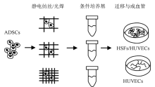
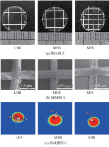
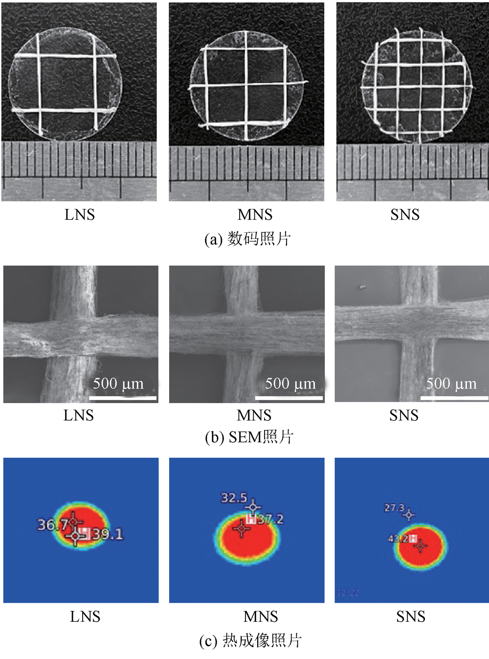
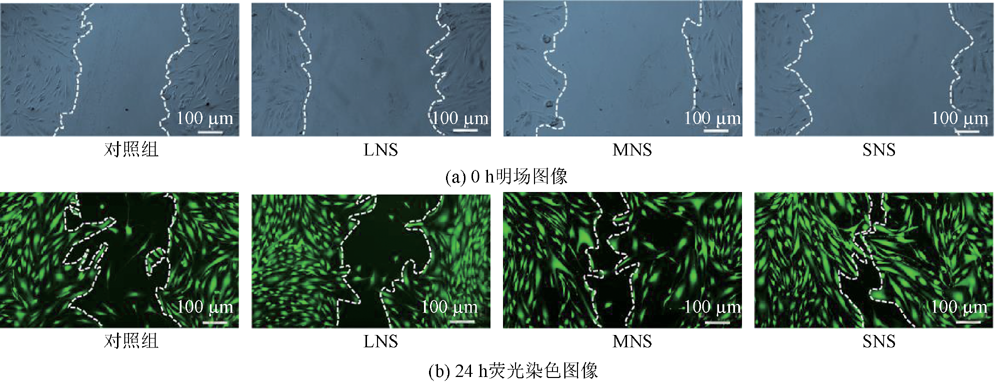
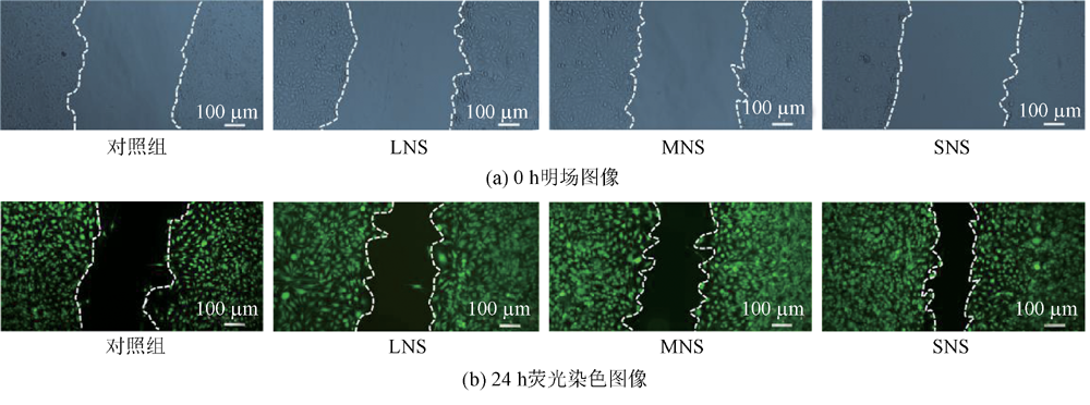
为探究纳米纤维纱线网格支架培养脂肪干细胞收集的条件培养基对创面修复过程中成纤维细胞和内皮细胞迁移以及血管化的调控作用,通过静电纺丝技术和光焊技术制备了大、中、小3种不同尺寸的纳米纤维纱线网格支架,并将其与脂肪干细胞共培养提取条件培养基,用于培养内皮细胞和成纤维细胞。使用扫描电子显微镜、数码相机和红外热像仪观察网格支架的微观、宏观结构和焊接温度,并通过细胞迁移实验和体外成血管实验评价3种条件培养基对细胞行为的调控作用。结果表明:制备的3种纳米纤维纱线网格支架微观形貌一致,纳米纤维纱线直径为(257.69 ± 36.87) μm,焊接温度为(39.83 ± 3.07) ℃;通过支架提取的条件培养基可促进成纤维细胞和内皮细胞的迁移及血管化,其中通过小网格支架提取的条件培养基对细胞迁移的促进效果更为明显,通过小网格和中网格支架提取的条件培养基对血管化的促进效果更明显。
中图分类号:
| [1] | LI J, YU F, CHEN G, et al. Moist-Retaining, Self-recoverable, bioadhesive, and transparent in situ forming hydrogels to accelerate wound healing[J]. ACS Applied Materials & Interfaces, 2020, 12(2): 2023-2038. |
| [2] | PEDRAM RAD Z, MOKHTARI J, ABBASI M. Fabrication and characterization of PCL/zein/gum arabic electrospun nanocomposite scaffold for skin tissue engineering[J]. Materials Science and Engineering: C, 2018, 93: 356-366. |
| [3] | 王曙东, 马倩, 王可, 等. 3D生物打印制备组织工程支架的研究进展[J]. 纺织学报, 2023, 44(3): 210-220. |
| WANG Shudong, MA Qian, WANG Ke, et al. Research progress in tissue engineering scaffolds by 3D bioprinting[J]. Journal of Textile Research, 2023, 44(3): 210-220. | |
| [4] | AN Y, LIN S, TAN X, et al. Exosomes from adipose-derived stem cells and application to skin wound hea-ling[J]. Cell Proliferation, 2021. DOI: 10.1111/jocd.13215. |
| [5] | GIZAW M, FAGLIE A, PIEPER M, et al. The role of electrospun fiber scaffolds in stem cell therapy for skin tissue regeneration[J]. Med One, 2019. DOI: 10.20900/mo.20190002. |
| [6] | HAO Z, QI W, SUN J, et al. Review: research progress of adipose-derived stem cells in the treatment of chronic wounds[J]. Frontiers in Chemistry, 2023. DOI: 10.3389/fchem.2023.1094693. |
| [7] | SCHNEIDER I, CALCAGNI M, BUSCHMANN J. Adipose-derived stem cells applied in skin diseases, wound healing and skin defects: a review[J]. Cytotherapy, 2023, 25(2): 105-119. |
| [8] | BORGESE M, BARONE L, ROSSI F, et al. Effect of nanostructured scaffold on human adipose-derived stem cells: outcome of in vitro experiments[J]. Nanomaterials, 2020. DOI: 10.3390/nano10091822. |
| [9] | LIN L, XU Y, LI Y, et al. Nanofibrous Wharton's jelly scaffold in combination with adipose-derived stem cells for cartilage engineering[J]. Materials & Design, 2020. DOI: 10.1016/j.matdes.2019.108216. |
| [10] | NING X, LIU N, SUN T, et al. Promotion of adipose stem cell transplantation using GelMA hydrogel reinforced by PLCL/ADM short nanofibers[J]. Biomedical Materials, 2023. DOI: 10.1088/1748-605x/acf551. |
| [11] | HUANG W, XIAO Y, SHI X. Construction of electrospun organic/inorganic hybrid nanofibers for drug delivery and tissue engineering applications[J]. Advanced Fiber Materials, 2019, 1(1): 32-45. |
| [12] | DONG Y, ZHENG Y, ZHANG K, et al. Electrospun nanofibrous materials for wound healing[J]. Advanced Fiber Materials, 2020, 2(4): 212-227. |
| [13] | NULTY J, FREEMAN F E, BROWE D C, et al. 3D bioprinting of prevascularised implants for the repair of critically-sized bone defects[J]. Acta Biomaterialia, 2021, 126: 154-169. |
| [14] | BACAKOVA L, ZARUBOVA J, TRAVNICKOVA M, et al. Stem cells: their source, potency and use in regenerative therapies with focus on adipose-derived stem cells - a review[J]. Biotechnology Advances, 2018, 36(4): 1111-1126. |
| [15] | YAO Q, COSME J G L, XU T, et al. Three dimensional electrospun PCL/PLA blend nanofibrous scaffolds with significantly improved stem cells osteogenic differentiation and cranial bone forma-tion[J]. Biomaterials, 2017, 115: 115-127. |
| [16] | CHEN S, LI R, LI X, et al. Electrospinning: An enabling nanotechnology platform for drug delivery and regenerative medicine[J]. Advanced Drug Delivery Reviews, 2018, 132: 188-213. |
| [17] | 付征, 穆齐峰, 张青松, 等. 胶体静电纺微纳米纤维的研究进展[J]. 纺织学报, 2023, 44(10): 196-204. |
| FU Zheng, MU Qifeng, ZHANG Qingsong, et al. Research progress in colloidal electrospun micro/nano fibers[J]. Journal of Textile Research, 2023, 44(10): 196-204. | |
| [18] | XIE J, MACEWAN M R, LI X, et al. Neurite outgrowth on nanofiber scaffolds with different orders, structures, and surface properties[J]. ACS Nano, 2009, 3(5): 1151-1159. |
| [19] | XUE J, WU T, DAI Y, et al. Electrospinning and electrospun nanofibers: methods, materials, and applications[J]. Chemical Reviews, 2019, 119(8): 5298-5415. |
| [20] | HEIDARI M, BAHRAMI H, RANJBAR-MOHAMMADI M. Fabrication, optimization and characterization of electrospun poly(caprolactone)/gelatin/graphene nanofibrous mats[J]. Materials Science and Engineering: C, 2017, 78: 218-229. |
| [21] | 贾姣, 郑作保, 吴昊, 等. 静电纺聚合物复合金属有机框架功能纳米纤维膜的研究进展[J]. 纺织学报, 2023, 44(6): 215-224. |
| JIA Jiao, ZHENG Zuobao, WU Hao, et al. Research progress in electrospinning functional nanofibers with metal-organic framework[J]. Journal of Textile Research, 2023, 44(6): 215-224. | |
| [22] | TASKIN M B, AHMAD T, WISTLICH L, et al. Bioactive electrospun fibers: fabrication strategies and a critical review of surface-sensitive characterization and quantification[J]. Chemical Reviews, 2021, 121(18): 11194-11237. |
| [23] | XUE J, PISIGNANO D, XIA Y. Maneuvering the migration and differentiation of stem cells with electrospun nanofibers[J]. Advanced Science, 2020, 7(15): 2000735. |
| [24] | LIANG R, ZHAO J, LI B, et al. Implantable and degradable antioxidant poly(ε-caprolactone)-lignin nanofiber membrane for effective osteoarthritis treat-ment[J]. Biomaterials, 2020. DOI: 10.1016/j.biomaterials.2019.119601. |
| [25] | SAWADKAR P, MOHANAKRISHNAN J, RAJASEKAR P, et al. A synergistic relationship between polycaprolactone and natural polymers enhances the physical properties and biological activity of sca-ffolds[J]. ACS Applied Materials & Interfaces, 2020, 12(12): 13587-13597. |
| [26] | FENG Z, ZHANG X, LIU N, et al. Promotion of neurite outgrowth and extension using injectable welded nanofibers[J]. Chemical Research in Chinese Universities, 2021, 37(3): 522-527. |
| [27] | WU T, XUE J, XIA Y. Engraving the surface of electrospun microfibers with nanoscale grooves promotes the outgrowth of neurites and the migration of schwann cells[J]. Angewandte Chemie International Edition, 2020, 59(36): 15626-15632. |
| [28] | LIU Y, ZHANG X, WANG Y, et al. Promoting neurite outgrowth and neural stem cell migration using aligned nanofibers decorated with protrusions and galectin-1 coating[J]. Chemical Communications, 2023, 59(72): 10753-10756. |
| [29] | KALIRAJAN C, BEHERA H, SELVARAJ V, et al. In vitro probing of oxidized inulin cross-linked collagen-ZrO2 hybrid scaffolds for tissue engineering applica-tions[J]. Carbohydrate Polymers, 2022. DOI: 10.1016/j.carbpol.2022.119458. |
| [30] | CHEN X, ZHANG L, CHAI W, et al. Hypoxic microenvironment reconstruction with synergistic biofunctional ions promotes diabetic wound healing[J]. Advanced Healthcare Materials, 2023. DOI: 10.1002/adhm.202301984. |
| [31] | DENG S, LEI T, CHEN H, et al. Metformin pre-treatment of stem cells from human exfoliated deciduous teeth promotes migration and angiogenesis of human umbilical vein endothelial cells for tissue enginee-ring[J]. Cytotherapy, 2022, 24(11): 1095-1104. |
| [32] | WU T, LI H, XUE J, et al. Photothermal welding, melting, and patterned expansion of nonwoven mats of polymer nanofibers for biomedical and printing applications[J]. Angewandte Chemie International Edition, 2019, 58(46): 16416-16421. |
| [33] | CHOI J K, JANG J H, JANG W H, et al. The effect of epidermal growth factor (EGF) conjugated with low-molecular-weight protamine (LMWP) on wound healing of the skin[J]. Biomaterials, 2012, 33(33): 8579-8590. |
| [34] | LIU Y, ZHU Z, PEI X, et al. ZIF-8-modified multifunctional bone-adhesive hydrogels promoting angiogenesis and osteogenesis for bone regenera-tion[J]. ACS Applied Materials & Interfaces, 2020, 12(33): 36978-36995. |
| [1] | 雷福旺, 冯其, 侯奥菡, 赵振鸿, 谭佳兆, 赵景, 王先锋. 聚偏氟乙烯-聚丙烯腈/SiO2单向导湿纤维膜的制备及其性能[J]. 纺织学报, 2024, 45(12): 1-8. |
| [2] | 刘霞, 吴改红, 闫子豪, 王彩柳. 智能相变调温聚乳酸纤维膜的制备及其性能[J]. 纺织学报, 2024, 45(12): 18-24. |
| [3] | 卢海龙, 于影, 左雨欣, 王浩然, 陈洪立, 汝欣. 取向增强抗CO2腐蚀纤维薄膜的制备及其性能[J]. 纺织学报, 2024, 45(12): 33-40. |
| [4] | 刘健, 董守骏, 王程皓, 刘泳汝, 潘山山, 尹兆松. 花瓣状多尖端静电纺丝喷头的电场模拟及优化[J]. 纺织学报, 2024, 45(10): 191-199. |
| [5] | 张佃平, 王昊, 林文峰, 王振秋. 多喷头纺丝装置的仿真与设计[J]. 纺织学报, 2024, 45(10): 200-207. |
| [6] | 蔺志浩, 房磊, 贾娇娇, 扈延龄, 房宽峻. 负载生长因子的微纳米纤维创面敷料的制备与应用研究进展[J]. 纺织学报, 2024, 45(09): 244-251. |
| [7] | 王浩然, 于影, 左雨欣, 顾志清, 卢海龙, 陈洪立, 柯俊. 聚乙烯亚胺/聚丙烯腈复合纤维薄膜的制备及其功能化应用[J]. 纺织学报, 2024, 45(09): 26-32. |
| [8] | 王清鹏, 张海艳, 王雨婷, 张涛, 赵燕. 聚环氧乙烷/Al2O3被动辐射降温膜的制备及其性能[J]. 纺织学报, 2024, 45(09): 33-41. |
| [9] | 钱洋, 张璐, 李晨阳, 王荣武. 静电纺海藻酸钠复合纳米纤维膜制备及其性能[J]. 纺织学报, 2024, 45(08): 18-25. |
| [10] | 刘嘉炜, 季东晓, 覃小红. 空气过滤用静电纺纳米纤维材料研究进展[J]. 纺织学报, 2024, 45(08): 35-43. |
| [11] | 刘德龙, 王红霞, 林童. 气流辅助的静电纺丝技术研究进展[J]. 纺织学报, 2024, 45(08): 44-53. |
| [12] | 杨硕, 赵朋举, 程春祖, 李晨暘, 程博闻. 非对称润湿性纤维复合膜的制备及其油水分离性能[J]. 纺织学报, 2024, 45(08): 10-17. |
| [13] | 王永政, 黄林涛, 宋付权. 石油沥青/聚丙烯腈静电纺碳纳米纤维的制备工艺优化及其性能[J]. 纺织学报, 2024, 45(08): 107-115. |
| [14] | 闫迪, 王雪芳, 谭文萍, 高国金, 明津法, 宁新. 富咪唑型多孔左旋聚乳酸纳米纤维膜制备及其双重净水性能[J]. 纺织学报, 2024, 45(08): 116-126. |
| [15] | 陈灿, 拖晓航, 王迎. 取向聚氨酯纳米纤维膜卷纱的制备及其力学性能[J]. 纺织学报, 2024, 45(08): 134-141. |
|
||
 京公网安备11010502044800号