纺织学报 ›› 2024, Vol. 45 ›› Issue (10): 224-231.doi: 10.13475/j.fzxb.20231006502
李蒙1, 戴梦男1, 俞杨销1, 王建南1,2( )
)
LI Meng1, DAI Mengnan1, YU Yangxiao1, WANG Jiannan1,2()
摘要:
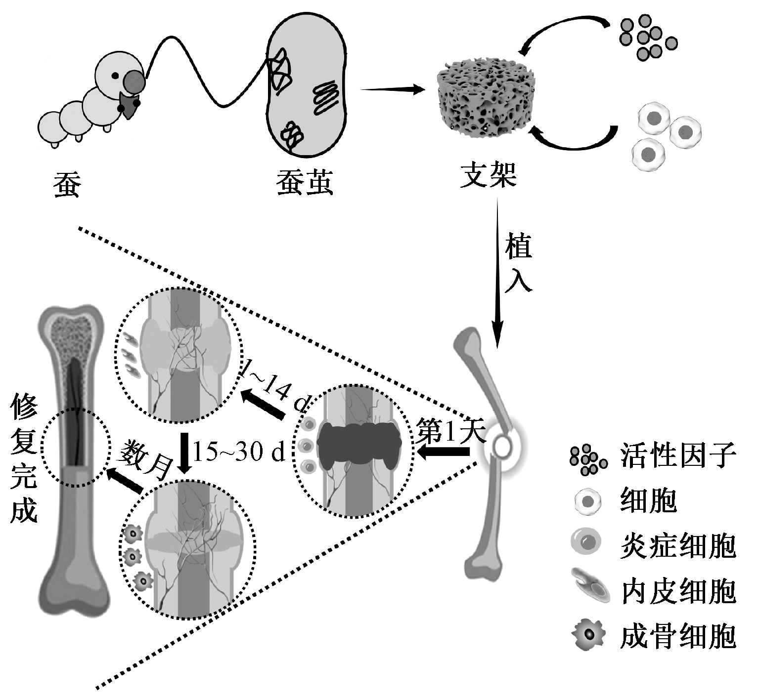
为拓展和促进丝素蛋白材料在骨再生领域中的应用,综述了近年来丝素蛋白骨修复材料的研究进展,简要概括了丝素蛋白骨修复材料的种类和制备方法,重点阐述了丝素蛋白骨修复材料的仿生设计以及力学性能和成骨性能的调控方法。分析发现:丝素蛋白基薄膜、水凝胶和多孔支架均能诱导骨再生;调控分子量、β-折叠结构或形成定向通道可有效提高材料的力学性能;与羟基磷灰石复合不仅能提高力学性能还可促进成骨分化;同时,负载细胞或生长因子可显著增加新生骨的体积,进而修复大尺寸骨缺损。最后,总结使用丝素蛋白修复骨缺损存在的问题,并指出丝素蛋白支架的仿生设计以及保持生长因子或种子细胞的活性仍是未来研究中考虑的重点。
中图分类号:
| [1] | XIE C, YE J, LIANG R J, et al. Advanced strategies of biomimetic tissue-engineered grafts for bone regeneration[J]. Advanced Healthcare Materials, 2021. DOI: 10.1002/adhm.202100408. |
| [2] | MA P F, WU W J, WEI Y, et al. Biomimetic gelatin/chitosan/polyvinyl alcohol/nano-hydroxyapatite scaffolds for bone tissue engineering[J]. Materials & Design, 2021. DOI: 10.1016/j.matdes.2021.109865. |
| [3] | JEYAKUMAR V, AMRAISH N, NICULESCU-MORSZA E, et al. Decellularized cartilage extracellular matrix incorporated silk fibroin hybrid scaffolds for endochondral ossification mediated bone regenera-tion[J]. International Journal of Molecular Sciences, 2021. DOI: 10.3390/ijms.22084055. |
| [4] | WU S L, LIU X M, YEUNG K W K, et al. Biomimetic porous scaffolds for bone tissue engineering[J]. Materials Science and Engineering R: Reports, 2014, 80(1): 1-36. |
| [5] |
MA H S, FENG C, CHANG J, et al. 3D-printed bioceramic scaffolds: from bone tissue engineering to tumor therapy[J]. Acta Biomaterialia, 2018, 79: 37-59.
doi: S1742-7061(18)30493-8 pmid: 30165201 |
| [6] |
WUBNEH A, TSEKOURA E K, AYRANCI C, et al. Current state of fabrication technologies and materials for bone tissue engineering[J]. Acta Biomaterialia, 2018, 80: 1-30.
doi: S1742-7061(18)30551-8 pmid: 30248515 |
| [7] | ZHANG D W, WU X W, CHEN J D, et al. The development of collagen based composite scaffolds for bone regeneration[J]. Bioactive Materials, 2017, 3(1): 129-138. |
| [8] |
BALAGANGADHARAN K, CHANDRAN S V, ARUMUGAM B, et al. Chitosan/nano-hydroxyapatite/nano-zirconium dioxide scaffolds with miR-590-5p for bone regeneration[J]. International Journal of Biological Macromolecules, 2018, 111: 953-958.
doi: S0141-8130(18)30019-9 pmid: 29415417 |
| [9] | 杨思敏, 王新卫. 自体骨移植修复骨缺损的临床研究进展[J]. 中国疗养医学, 2019, 9: 945-948. |
| YANG Simin, WANG Xinwei. Clinical research progress of autogenous bone transplantation for repairing bone defect[J]. Chinese Journal of Convalescent Medicine, 2019, 9: 945-948. | |
| [10] |
ROBERTS T T, ROSENBAUM A J. Bone grafts, bone substitutes and orthobiologics: the bridge between basic science and clinical advancements in fracture healing[J]. Organogenesis, 2012, 8(4): 114-124.
doi: 10.4161/org.23306 pmid: 23247591 |
| [11] | 胡居正, 石展英, 杨成志. 同种异体骨移植治疗骨缺损的应用研究进展[J]. 基层医学论坛, 2017, 19: 2570-2572. |
| HU Juzheng, SHI Zhanying, YANG Chengzhi. Application and research progress of allogeneic bone transplantation for the treatment of bone defect[J]. The Medical Forum, 2017, 19: 2570-2572. | |
| [12] |
东家慧, 谭丽丽, 杨柯. 可降解镁基金属骨缺损修复材料的研究探索[J]. 金属学报, 2017, 53(10): 1197-1206.
doi: 10.11900/0412.1961.2017.00279 |
|
DONG Jiahui, TAN Lili, YANG Ke. Research of biodegradable mg-based metals as bone graft substitutes[J]. Acta Metallurgica Sinica, 2017, 53(10): 1197-1206.
doi: 10.11900/0412.1961.2017.00279 |
|
| [13] | 韦章澳, 徐凌寒, 吴子辰, 等. 无机非金属人工骨修复材料的体内应用[J]. 中国组织工程研究, 2022, 16: 2706-2712. |
| WEI Zhang'ao, XU Linghan, WU Zichen, et al. Application of inorganic nonmetallic artificial bone materials in vivo[J]. Chinese Journal of Tissue Engineering Research, 2022, 16: 2706-2712. | |
| [14] | ZHAO Y, ZHAO S N, MA Z X, et al. Chitosan-based scaffolds for facilitated endogenous bone regenera-tion[J]. Pharmaceuticals, 2022. DOI: 10.3390/ph15081023. |
| [15] | GAO H C, GE K K, XU Y Q, et al. Controlled release of minocycline in hydroxyapatite/chitosan composite for periodontal bone defect repair[J]. Dental Materials Journal, 2022, 41(3): 346-352. |
| [16] | CHENG Y, CHENG G, XIE C Y. Biomimetic silk fibroin hydrogels strengthened by silica nanoparticles distributed nanofibers facilitate bone repair[J]. Advanced Healthcare Materials, 2021. DOI:10.1002/adhm.202001646. |
| [17] | WANG C L, MENG C Y, ZHANG Z, et al. 3D printing of polycaprolactone/bioactive glass composite scaffolds for in situ bone repair[J]. Ceramics International, 2022, 48(6): 7491-7499. |
| [18] | LI B, QU M Y, YANG H C, et al. Biomimetic mineralization of poly(L-lactic acid) nanofibrous microspheres for bone regeneration[J]. Materials Today Communications, 2022. DOI: 10.1016/j.mtcomm.2022.104682. |
| [19] | CARMAGNOLA I, NARDO T, GENTILE P. Poly(lactic acid)-based blends with tailored physicochemical properties for tissue engineering application: a case study[J]. International Journal of Polymeric Materials and Polymeric Biomaterials, 2015, 64(2): 90-98. |
| [20] | 罗元泽, 戴梦男, 李蒙, 等. 丝素蛋白基药物载体的应用研究进展[J]. 纺织学报, 2023, 44(9): 213-222. |
| LUO Yuanze, DAI Mengnan, LI Meng, et al. Application of silk fibroin-based biomaterials for drug delivery[J]. Journal of Textile Research, 2023, 44(9): 213-222. | |
| [21] | 黄利. 丝素蛋白仿生组织工程支架的成型、结构与性能研究[D]. 上海: 东华大学, 2020: 7-8. |
| HUANG Li. Preparation, structures and properties of silk fibroin based biomimetic tissue engineering sca-ffolds[D]. Shanghai: Donghua University, 2020: 7-8. | |
| [22] | 于成龙, 关国平, 余劭婷, 等. 丝素分子的构象转变及自组装行为[J]. 生物医学工程学进展, 2017, 38(3): 159-163. |
| YU Chenglong, GUAN Guoping, YU Shaoting, et al. Conformation transition and self-assembly of silk fibroin[J]. Progress in Biomedical Engineering, 2017, 38(3): 159-163. | |
| [23] |
MELKE J, MIDHA S, GHOSH S, et al. Silk fibroin as biomaterial for bone tissue engineering[J]. Acta Biomaterialia, 2016, 31: 1-16.
doi: S1742-7061(15)30098-2 pmid: 26360593 |
| [24] | 陈智洋, 叶军, 王洪亮, 等. 基于丝素蛋白的纳米粒药物递送系统研究进展[J]. 药学学报, 2022, 6: 1792-1800. |
| CHEN Zhiyang, YE Jun, WANG Hongliang, et al. Research progress of silk fibroin-based nanoparticulate drug delivery systems[J]. Acta Pharmaceutica Sinica, 2022, 6: 1792-1800. | |
| [25] | 高舒颖, 徐莹颖, 李曦. 基于丝素蛋白的新型药物递送系统研究进展[J]. 药学与临床研究, 2021, 5: 371-376. |
| GAO Shuying, XU Yingying, LI Xi. Advances in silk fibroin-based novel drug delivery systems[J]. Pharmaceutical and Clinical Research, 2021, 5: 371-376. | |
| [26] |
LIU L, YU F, LI L, et al. Bone marrow stromal cells stimulated by strontium-substituted calcium silicate ceramics: release of exosomal miR-146a regulates osteogenesis and angiogenesis[J]. Acta Biomaterialia, 2021, 119: 444-457.
doi: 10.1016/j.actbio.2020.10.038 pmid: 33129987 |
| [27] | DONG Y, LIU Y, CHEN Y, et al. Spatiotemporal regulation of endogenous MSCs using a functional injectable hydrogel system for cartilage regeneration[J]. NPG Asia Materials, 2021. DOI: 10.1038/s41427-021-00339-3. |
| [28] | FAN Z H, LIU H X, SHI S L, et al. Anisotropic silk nanofiber layers as regulators of angiogenesis for optimized bone regeneration[J]. Materials Today Bio, 2022. DOI: 10.1016/j.mtbio.2022.100283. |
| [29] | 千建峰, 蔡丽慧, 亓卫东, 等. 不同孔径丝素蛋白支架体内降解观察[J]. 中国生物医学工程学报, 2016, 4: 507-511. |
| QIAN Jianfeng, CAI Lihui, QI Weidong, et al. Degradation behaviors of silk fibroin scaffolds with different pore sizes in vivo[J]. Chinese Journal of Biomedical Engineering, 2016, 4: 507-511. | |
| [30] | WANG J N, LIU Z W, YANG Y X, et al. Enzymatic degradation behavior of silk fibroin fiber treated by gamma-ray irradiation[J]. Textile Research Journal, 2012, 82(17): 1799-1805. |
| [31] |
SENGUPTA S, PARK S H, SEOK G E, et al. Quantifying osteogenic cell degradation of silk biomaterials[J]. Biomacromolecules, 2010, 11(12): 3592-3599.
doi: 10.1021/bm101054q pmid: 21105641 |
| [32] | WANG L P, LUO Z W, ZHANG Q, et al. Effect of degumming methods on the degradation behavior of silk fibroin biomaterials[J]. Fibers and Polymers, 2019, 20(1): 45-50. |
| [33] | LI M, TIAN W, YU Y X, et al. Effect of degumming degree on the structure and tensile properties of RSF/RSS composite films prepared by one-step extraction[J]. Scientific Reports, 2023. DOI: 10.1038/s41598-023-33844-2. |
| [34] | WANG Q, XU J X, JIN H M, et al. Artificial periosteum in bone defect repair: a review[J]. Chinese Chemical Letters, 2017, 28 (9): 1801-1807. |
| [35] | YANG G J, LIU H M, CUI Y, et al. Bioinspired membrane provides periosteum-mimetic microenvironment for accelerating vascularized bone regeneration[J]. Biomaterials, 2021. DOI: 10.1016/j.biomaterials.2020.120561. |
| [36] | LI M, TIAN W, ZHANG Y, et al. Enhanced silk fibroin/sericin composite film: preparation, mechanical properties and mineralization activity[J]. Polymers, 2022. DOI: 10.3390/polym14122466. |
| [37] |
ZHENG X, KE X, YU P, et al. A facile strategy to construct silk fibroin based GTR membranes with appropriate mechanical performance and enhanced osteogenic capacity[J]. Journal of Materials Chemistry B, 2020, 8(45): 10407-10415.
doi: 10.1039/d0tb01962c pmid: 33112359 |
| [38] |
REAKASAME S, BOCCACCINI A R. Oxidized alginate-based hydrogels for tissue engineering applications: a review[J]. Biomacromolecules, 2018, 19(1): 3-21.
doi: 10.1021/acs.biomac.7b01331 pmid: 29172448 |
| [39] | ZHANG X Y, XIAO L Y, DING Z Z, et al. Engineered tough silk hydrogels through assembling β-sheet rich nanofibers based on a solvent replacement strategy[J]. ACS Nano, 2022, 16: 10209-10218. |
| [40] | SRISAWASDI T, PETCHAROEN K, SIRIVAT A, et al. Electromechanical response of silk fibroin hydrogel and conductive polycarbazole/silk fibroin hydrogel composites as actuator material[J]. Materials Science & Engineering C: Materials For Biological Applications, 2015, 56: 1-8. |
| [41] | ELLIOTT W H, BONANI W, MANIGLIO D, et al. Silk hydrogels of tunable structure and viscoelastic properties using different chronological orders of genipin and physical cross-linking[J]. ACS Applied Materials & Interfaces, 2015, 7(22): 12099-12108. |
| [42] |
MCGILL M, COBURN J M, PARTLOW B P, et al. Molecular and macro-scale analysis of enzyme-crosslinked silk hydrogels for rational biomaterial design[J]. Acta Biomaterialia, 2017, 63: 76-84.
doi: S1742-7061(17)30583-4 pmid: 28919509 |
| [43] | WANG Y, YANG Z Y, CHEN X, et al. Silk fibroin hydrogel membranes prepared by a sequential cross-linking strategy for guided bone regeneration[J]. Journal of the Mechanical Behavior of Biomedical Materials, 2023. DOI: 10.1016/j.jmbbm.2023.106133. |
| [44] | ZHAN J L, SUN X D, CUI F Z, et al. Preparation of 3-D porous fibiroin scaffolds by freeze drying with treatment of methanol solutions[J]. Chinese Science Bulletin, 2007, 52(13): 1791-1795. |
| [45] |
OLIVEIRA A L, SUN L, KIM H J, et al. Aligned silk-based 3-D architectures for contact guidance in tissue engineering[J]. Acta Biomaterialia, 2012, 8: 1530-1542.
doi: 10.1016/j.actbio.2011.12.015 pmid: 22202909 |
| [46] |
FAN L P, LI J L, CAI Z X, et al. Creating biomimetic anisotropic architectures with co-aligned nanofibers and macrochannels by manipulating ice crystallization[J]. ACS Nano, 2018, 12: 5780-5790.
doi: 10.1021/acsnano.8b01648 pmid: 29846058 |
| [47] | DEININGER C, WAGNER A, HEIMEL P, et al. Enhanced BMP-2-mediated bone repair using an anisotropic silk fibroin scaffold coated with bone-like apatite[J]. International Journal of Molecular Sciences, 2022. DOI: 10.3390/ijms23010283. |
| [48] |
PARK H J, LEE O J, LEE M C, et al. Fabrication of 3D porous silk scaffolds by particulate (salt/sucrose) leaching for bone tissue reconstruction[J]. International Journal of Biological Macromolecules, 2015, 78: 215-223.
doi: 10.1016/j.ijbiomac.2015.03.064 pmid: 25849999 |
| [49] |
CORREIA C, BHUMIRATANA S, YAN L P, et al. Development of silk-based scaffolds for tissue engineering of bone from human adipose-derived stem cells[J]. Acta Biomaterialia, 2012, 8(7): 2483-2492.
doi: 10.1016/j.actbio.2012.03.019 pmid: 22421311 |
| [50] | 耿亚楠, 赵梦露, 姚响, 等. 强韧支架用丝素蛋白基生物墨水及其3D打印支架模拟软件的开发[J]. 功能高分子学报, 2023(2): 107-116. |
| GENG Yanan, ZHAO Menglu, YAO Xiang, et al. Development of silk fibroin-based bio-inks for strong scaffolds and 3D printing simulation software for scaffolds[J]. Journal of Functional Polymers, 2023(2): 107-116. | |
| [51] | WANG Q, HAN G, YAN S, et al. 3D printing of silk fibroin for biomedical applications[J]. Materials, 2019. DOI: 10.3390/ma12030504. |
| [52] | SUN M Y, CHI G F, XU J J, et al. Extracellular matrix stiffness controls osteogenic differentiation of mesenchymal stem cells mediated by integrin α5[J]. Stem Cell Research & Therapy, 2018. DOI: 10.1186/s13287-018-0798-0. |
| [53] | YAN L P, SILVA-CORREIA J, OLIVEIRA M B, et al. Bilayered silk/silk-nanoCaP scaffolds for osteochondral tissue engineering: in vitro and in vivo assessment of biological performance[J]. Acta Biomaterialia, 2015, 12: 227-241. |
| [54] |
YAN Z, CHEN W B, JIN W H, et al. An interference screw made using a silk fibroin-based bulk material with high content of hydroxyapatite for anterior cruciate ligament reconstruction in a rabbit model[J]. Journal of Materials Chemistry B, 2021, 9(26): 5352-5364.
doi: 10.1039/d1tb01006a pmid: 34152356 |
| [55] | JIN S, FU X X, ZENG W A, et al. Chopped fibers and nano-hydroxyapatite enhanced silk fibroin porous hybrid scaffolds for bone augmentation[J]. Journal of Materials Chemistry B, 2023, 11(7): 1557-1567. |
| [56] |
YAO Y K, GUAN D Q, ZHANG C K, et al. Silkworm spinning inspired 3D printing toward a high strength scaffold for bone regeneration[J]. Journal of Materials Chemistry B, 2022, 10(36): 6946-6957.
doi: 10.1039/d2tb01161a pmid: 36069158 |
| [57] | WU J N, CAO L Y, LIU Y, et al. Functionalization of silk fibroin electrospun scaffolds via bmsc affinity peptide grafting through oxidative self-polymerization of dopamine for bone regeneration[J]. ACS Applied Materials & Interfaces, 2019, 11(9): 8878-8895. |
| [58] | SARTIKA D, WANG C H, WANG D H, et al. Human adipose-derived mesenchymal stem cells-incorporated silk fibroin as a potential bio-scaffold in guiding bone regeneration[J]. Polymers, 2020. DOI: 10.3390/polym12040853. |
| [59] | YU X, WAN Q L, YE X L, et al. Cellular hypoxia promotes osteogenic differentiation of mesenchymal stem cells and bone defect healing via STAT3 signaling[J]. Cellular & Molecular Biology Letters, 2019. DOI: 10.1186/s11658-019-0191-8. |
| [60] | SUN J C, LI L, XING F, et al. Graphene oxide-modified silk fibroin/nanohydroxyapatite scaffold loaded with urine-derived stem cells for immunomodulation and bone regeneration[J]. Stem Cell Research & Therapy, 2021. DOI: 10.1186/s13287-021-02634-w. |
| [61] | WANG L, LIAN J, XIA Y J, et al. A study on in vitro and in vivo bioactivity of silk fibroin/nano-hydroxyapatite/graphene oxide composite scaffolds with directional channels[J]. Colloids and Surfaces A: Physicochemical and Engineering Aspects, 2022. DOI: 10.1016/j.colsurfa.2022.129886. |
| [1] | 王宇航 谭晶 李好义 徐锦龙 杨卫民. 纳米纤维纱线静电纺制备技术研究进展[J]. , 2024, 45(11): 0-0. |
| [2] | 刘婷, 闫涛, 潘志娟. 香蕉茎秆纤维/抗菌纤维混纺纱的制备及其性能[J]. 纺织学报, 2024, 45(10): 48-54. |
| [3] | 王勃翔, 徐航丹, 李佳, 林杰, 程德红, 路艳华. 柞蚕丝素纳米纤维温敏复合膜制备及其生物相容性[J]. 纺织学报, 2024, 45(09): 18-25. |
| [4] | 徐豫松, 周杰, 甘佳怡, 张涛, 张先明. 含磷氮水性聚氨酯的制备及其在涤纶织物阻燃整理中应用[J]. 纺织学报, 2024, 45(07): 112-120. |
| [5] | 于承浩, 王元非, 于腾波, 吴桐. 热致自卷曲左旋聚乳酸/聚乳酸-羟基乙酸共聚物纳米纤维血管支架制备及其性能[J]. 纺织学报, 2024, 45(07): 18-23. |
| [6] | 刘术, 侯腾, 周乐乐, 李祥龙, 杨斌. 桑蚕的强制牵伸抽丝及其纤维性能[J]. 纺织学报, 2024, 45(06): 11-15. |
| [7] | 黄晴, 苏振岳, 周一帆, 刘青松, 李懿, 赵萍, 王鑫. 饲料与桑叶饲喂的家蚕蚕丝品质分析[J]. 纺织学报, 2024, 45(05): 1-9. |
| [8] | 丁彩红, 顾馨, 卢晨雨. 血管支架预制件的六角形三维虚拟编织[J]. 纺织学报, 2024, 45(03): 65-73. |
| [9] | 谷金峻, 魏春艳, 郭紫阳, 吕丽华, 白晋, 赵航慧妍. 棉秆皮微晶纤维素/改性氧化石墨烯阻燃纤维的制备及其性能[J]. 纺织学报, 2024, 45(01): 39-47. |
| [10] | 雷彩虹, 俞林双, 金万慧, 朱海霖, 陈建勇. 丝素蛋白/壳聚糖复合纤维膜的制备与应用[J]. 纺织学报, 2023, 44(11): 19-26. |
| [11] | 陈美玉, 李立凤, 董侠. 长碳链聚酰胺1012纤维在不同温度下的力学性能[J]. 纺织学报, 2023, 44(11): 9-18. |
| [12] | 张子凡, 李鹏飞, 王建南, 许建梅. 丝素蛋白载药纳米粒的研究进展[J]. 纺织学报, 2023, 44(10): 205-213. |
| [13] | 罗元泽, 戴梦男, 李蒙, 俞杨销, 王建南. 丝素蛋白基药物载体的应用研究进展[J]. 纺织学报, 2023, 44(09): 213-222. |
| [14] | 张颖, 宋明根, 姬洪, 陈康, 张先明. 热定形工艺对高强型聚酯工业丝结构性能的影响[J]. 纺织学报, 2023, 44(09): 43-51. |
| [15] | 孙明涛, 陈成玉, 闫伟霞, 曹珊珊, 韩克清. 针刺加固频率对黄麻纤维/聚乳酸短纤复合板性能的影响[J]. 纺织学报, 2023, 44(09): 91-98. |
|
||
 京公网安备11010502044800号