纺织学报 ›› 2025, Vol. 46 ›› Issue (01): 206-216.doi: 10.13475/j.fzxb.20240103502
杨柳1,2, 杜磊1( ), 徐淮中2
YANG Liu1,2, DU Lei1(), XU Huaizhong2
摘要:
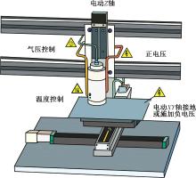
熔体近场直写(MEW)作为一种新兴的增材制造技术,可在微米尺度下实现纤维支架结构的精准构筑。为进一步推动该技术在组织工程领域的应用,综述了近年来国内外MEW技术的研究进展。针对MEW设备组成,详解了各模块的工作原理和设计思路;针对MEW射流调控,阐述了射流稳定及沉积过程中的成形机制与控制机制;针对MEW技术的应用场景,讨论了不同结构支架的构筑方法及其构效关系;此外,还总结了MEW技术与其它技术结合的典型范例;最后,展望了MEW技术的发展方向,以期为其在多领域交叉融合发展提供理论支撑和应用参考。
中图分类号:
| [1] | BROWN Toby D, DALTON Paul D, HUTMACHER Dietmar W. Direct writing by way of melt electro-spinning[J]. Advanced Materials, 2011, 23(47): 5651-5657. |
| [2] | EICHHOLZ Kian F, GONÇALVES Inês, BARCELÓ Xavier, et al. How to design, develop and build a fully-integrated melt electrowriting 3D printer[J]. Additive Manufacturing, 2022. DOI: 10.1016/j.addma.2022.102998. |
| [3] | LU Huali, SUN Yue, CHEN Yufei, et al. The effects of voltage configurations on print accuracy in melt electrowriting[J]. Materials Letters, 2023. DOI: 10.1016/j.matlet.2022.133738. |
| [4] | TOURLOMOUSIS Filippos, DING Houzhu, KALYON Dilhan M, et al. Melt electrospinning writing process guided by a ″Printability Number″[J]. Journal of Manufacturing Science and Engineering, 2017. DOI: 10.1115/1.4036348. |
| [5] | HE Jiankang, XIA Peng, LI Dichen. Development of melt electrohydrodynamic 3D printing for complex microscale poly (ε-caprolactone) scaffolds[J]. Biofabrication, 2016. DOI: 10.1088/1758-5090/8/3/035008. |
| [6] | ZHANG Fucheng, CAO Kai, ZAERI Ahmadreza, et al. Effects of scaffold design parameters on the printing accuracy for melt electrowriting[J]. Journal of Manufacturing Processes, 2022, 81: 177-190. |
| [7] | HOCHLEITNER Gernot, HÜMMER Julia Franziska, LUXENHOFER Robert, et al. High definition fibrous poly(2-ethyl-2-oxazoline) scaffolds through melt electrospinning writing[J]. Polymer, 2014, 55(20): 5017-5023. |
| [8] | MIESZCZANEK Pawel, ROBINSON Thomas M, DALTON Paul D, et al. Convergence of machine vision and melt electrowriting[J]. Advanced Materials, 2021. DOI: 10.1002/adma.202100519. |
| [9] | HOCHLEITNER Gernot, YOUSSEF Almoatazbellah, HRYNEVICH Andrei, et al. Fibre pulsing during melt electrospinning writing[J]. BioNanoMaterials, 2016, 17(3/4): 159-171. |
| [10] | NADERNEZHAD Ali, RYMA Matthias, GENÇ Hatice, et al. Melt electrowriting of isomalt for high-resolution templating of embedded microchannels[J]. Advanced Materials Technologies, 2021. DOI: 10.1002/admt.202100221. |
| [11] | CAO Kai, ZHANG Fucheng, WANG Bijun, et al. Analytical interpretation of microscale fiber deviation in designing for polymer melt electrohydrodynamic-based additive manufacturing[J]. Additive Manufacturing, 2022. DOI: 10.1016/j.addma.2022.103035. |
| [12] | XU Huaizhong, LIASHENKO Ievgenii, LUCCHETTI Agnese, et al. Designing with circular arc toolpaths to increase the complexity of melt electrowriting[J]. Advanced Materials Technologies, 2022. DOI: 10.1002/admt.202101676. |
| [13] | ROBINSON Thomas M, HUTMACHER Dietmar W, DALTON Paul D. The next frontier in melt electrospinning: taming the jet[J]. Advanced Functional Materials, 2019. DOI: 10.1002/adfm.201904664. |
| [14] | SU Yingchun, ZHANG Zhongyang, WAN Yilin, et al. A hierarchically ordered compacted coil scaffold for tissue regeneration[J]. NPG Asia Materials, 2020. DOI: 10.1038/s41427-020-0234-7. |
| [15] | KIM Jaehyeon, BAKIRCI Ezgi, O'NEILL Kelly L, et al. Fiber bridging during melt electrowriting of poly (ε-caprolactone) and the influence of fiber diameter and wall height[J]. Macromolecular Materials and Engineering, 2021. DOI: 10.1002/mame.202000685. |
| [16] | DU Lei, YANG Liu, LU Huali, et al. Additive manufacturing of ultrahigh-resolution poly(ε-caprolactone) scaffolds using melt electrowriting[J]. Polymer, 2024. DOI: 10.1016/j.polymer.2024.127028. |
| [17] | HE Jiankang, HAO Guanzhe, MENG Zijie, et al. Expanding melt-based electrohydrodynamic printing of highly-ordered microfibrous architectures to cm-height via in situ charge neutralization[J]. Advanced Materials Technologies, 2022. DOI: 10.1002/admt.202101197. |
| [18] | LIASHENKO Ievgenii, HRYNEVICH Andrei, DALTON Paul D. Designing outside the box: unlocking the geometric freedom of melt electrowriting using microscale layer shifting[J]. Advanced Materials, 2020. DOI: 10.1002/adma.202001874. |
| [19] | MENG Jie, BOSCHETTO Francesco, YAGI Shinichi, et al. Design and manufacturing of 3D high-precision micro-fibrous poly (L-lactic acid) scaffold using melt electrowriting technique for bone tissue engineering[J]. Materials and Design, 2021. DOI: 10.1016/j.matdes.2021.110063. |
| [20] | NGUYEN Nhat Tung, KIM Jeong Hwa, JEONG Young Hun. Identification of sagging in melt-electrospinning of microfiber scaffolds[J]. Materials Science and Engineering: C, 2019. DOI: 10.1016/j.msec.2019.109785. |
| [21] | HRYNEVICH Andrei, ACHENBACH Pascal, JUNGST Tomasz, et al. Design of suspended melt electrowritten fiber arrays for Schwann cell migration and neurite outgrowth[J]. Macromolecular Bioscience, 2021. DOI: 10.1002/mabi.202000439. |
| [22] | BERTLEIN Sarah, HIKIMOTO Daichi, HOCHLEITNER Gernot, et al. Development of endothelial cell networks in 3D tissues by combination of melt electrospinning writing with cell-accumulation technology[J]. Small, 2018. DOI: 10.1002/smll.201701521. |
| [23] | CASTILHO Miguel, FEYEN Dries, FLANDES-IPARRAGUIRRE María, et al. Melt electrospinning writing of poly-hydroxymethylglycolide-co-ε-caprolactone-based scaffolds for cardiac tissue engineering[J]. Advanced Healthcare Materials, 2017. DOI: 10.1002/adhm.201700311. |
| [24] | CASTILHO Miguel, VAN MIL Alain, MAHER Malachy, et al. Melt electrowriting allows tailored microstructural and mechanical design of scaffolds to advance functional human myocardial tissue formation[J]. Advanced Functional Materials, 2018. DOI: 10.1002/adfm.201803151. |
| [25] | CEDILLO-SERVIN Gerardo, DAHRI Ouafa, MENESES João, et al. 3D printed magneto-active microfiber scaffolds for remote stimulation and guided organization of 3D in vitro skeletal muscle models[J]. Small, 2023. DOI: 10.1002/smll.202307178. |
| [26] | MONDADORI Carlotta, CHANDRAKAR Amit, LOPA Silvia, et al. Assessing the response of human primary macrophages to defined fibrous architectures fabricated by melt electrowriting[J]. Bioactive Materials, 2023(21): 209-222. |
| [27] | LI Sicheng, HUANG Jinjian, XU Ziyan, et al. Melt electrowriting-printed peritoneal scaffold prevents peritoneal adhesion and facilitates peritoneal repair[J]. International Journal of Bioprinting, 2023. DOI: 10.18063/ijb.682. |
| [28] | DAGHRERY Arwa, FERREIRA Jessica A, DE SOUZA Araújo Isaac J, et al. A highly ordered, nanostructured fluorinated CaP-coated melt electrowritten scaffold for periodontal tissue regeneration[J]. Advanced Healthcare Materials, 2021. DOI: 10.1002/adhm.202101152. |
| [29] | BARCELÓ Xavier, EICHHOLZ Kian F, GONÇALVES Inês F, et al. Bioprinting of structurally organized meniscal tissue within anisotropic melt electrowritten scaffolds[J]. Acta Biomaterialia, 2023, 158: 216-227. |
| [30] | BAS Onur, D'ANGELLA Davide, BALDWIN Jeremy G, et al. An integrated design, material, and fabrication platform for engineering biomechanically and biologically functional soft tissues[J]. ACS Applied Materials and Interfaces, 2017, 9(35): 29430-29437. |
| [31] | SAIDY Navid T, WOLF Frederic, BAS Onur, et al. Biologically inspired scaffolds for heart valve tissue engineering via melt electrowriting[J]. Small, 2019. DOI: 10.1002/smll.201900873. |
| [32] | OLVERA Dinorath, SOHRABI MOLINA Mina, HENDY Gillian, et al. Electroconductive melt electrowritten patches matching the mechanical anisotropy of human myocardium[J]. Advanced Functional Materials, 2020. DOI: 10.1002/adfm.201909880. |
| [33] | SHAFIEE Abbas, CAVALCANTI Amanda S, SAIDY Navid T, et al. Convergence of 3D printed biomimetic wound dressings and adult stem cell therapy[J]. Biomaterials, 2021. DOI: 10.1016/j.biomaterials.2020.120558. |
| [34] | MATHEW Asha, DEVLIN Brenna L, SINGH Dilpreet, et al. Improving infection resistance in tissue engineered scaffolds for tensile applications using vancomycin-embedded melt electrowritten scaffolds[J]. Macromolecular Materials and Engineering, 2023. DOI: 10.1002/mame.202300168. |
| [35] | YANG Liu, LOU Yi, ZHANG Guoping, et al. Hybrid manufacturing of highly stretchable functionalized membrane for joint wound treatment[J]. Colloids and Surfaces A: Physicochemical and Engineering Aspects, 2024. DOI: 10.1016/j.colsurfa.2023.132655. |
| [36] | JIN Yuan, GAO Qing, XIE Chaoqi, et al. Fabrication of heterogeneous scaffolds using melt electrospinning writing: design and optimization[J]. Materials and Design, 2020. DOI: 10.1016/j.matdes.2019.108274. |
| [37] | XIE Chaoqi, GAO Qing, WANG Peng, et al. Structure-induced cell growth by 3D printing of heterogeneous scaffolds with ultrafine fibers[J]. Materials and Design, 2019. DOI: 10.1016/j.matdes.2019.108092. |
| [38] | VERNON Michael J, LU Jason, PADMAN Benjamin, et al. Engineering heart valve interfaces using melt electrowriting: biomimetic design strategies from multi-modal imaging[J]. Advanced Healthcare Materials, 2022. DOI: 10.1002/adhm.202201028. |
| [39] | ABBASI Naghmeh, LEE Ryan S B, IVANOVSKI Saso, et al. In vivo bone regeneration assessment of offset and gradient melt electrowritten (MEW) PCL scaffolds[J]. Biomaterials Research, 2020. DOI: 10.1186/s40824-020-00196-1. |
| [40] | GOLAFSHAN Nasim, CASTILHO Miguel, DAGHRERY Arwa, et al. Composite graded melt electrowritten scaffolds for regeneration of the periodontal ligament-to-bone interface[J]. ACS Applied Materials and Interfaces, 2023, 15(10): 12735-12749. |
| [41] | PAXTON Naomi C, LUPOSCHAINSKY Simon, REIZABAL Ander, et al. Manufacture of biomimetic auricular surgical implants using 3D printed high density polyethylene microfibers[J]. Advanced Materials Technologies, 2023. DOI: 10.1002/admt.202301190. |
| [42] | BROWN Toby D, SLOTOSCH Anna, THIBAUDEAU Laure, et al. Design and fabrication of tubular scaffolds via direct writing in a melt electrospinning mode[J]. Biointerphases, 2012. DOI: 10.1007/s13758-011-0013-7. |
| [43] | MCCOLL Erin, GROLL Jürgen, JUNGST Tomasz, et al. Design and fabrication of melt electrowritten tubes using intuitive software[J]. Materials and Design, 2018, 155: 46-58. |
| [44] | ZHANG Fucheng, CAO Kai, ZAERI Ahmadreza, et al. Design, fabrication, and characterization of tubular scaffolds by way of a melt electrowriting process[J]. Additive Manufacturing, 2023. DOI: 10.1016/j.addma.2022.103383. |
| [45] | PAXTON Naomi C, DALEY Ryan, FORRESTAL David P, et al. Auxetic tubular scaffolds via melt electrowriting[J]. Materials and Design, 2020. DOI:10.1016/j.matdes.2020.108787. |
| [46] | MCCOSKER Audrey B, SNOWDON Mikayla E, LAMONT Riki, et al. Exploiting nonlinear fiber patterning to control tubular scaffold mechanical behavior[J]. Advanced Materials Technologies, 2022. DOI: 10.1002/admt.202200259. |
| [47] | SOMSZOR Katarzyna, BAS Onur, KARIMI Fatemeh, et al. Personalized, mechanically strong, and biodegradable coronary artery stents via melt electrowriting[J]. ACS Macro Letters, 2020, 9(12): 1732-1739. |
| [48] | SAIDY Navid T, SHABAB Tara, BAS Onur, et al. Melt electrowriting of complex 3D anatomically relevant scaffolds[J]. Frontiers in Bioengineering and Biotechnology, 2020. DOI: 10.3389/fbioe.2020.00793. |
| [49] | SAIDY Navid Toosi, FERNÁNDEZ-COLINO Alicia, HEIDARI Behzad Shiroud, et al. Spatially heterogeneous tubular scaffolds for in situ heart valve tissue engineering using melt electrowriting[J]. Advanced Functional Materials, 2022. DOI: 10.1002/adfm.202110716. |
| [50] | WEEKES Angus, WEHR Gabrielle, PINTO Nigel, et al. Highly compliant biomimetic scaffolds for small diameter tissue-engineered vascular grafts (TEVGs) produced via melt electrowriting (MEW)[J]. Biofabrication, 2023. DOI: 10.1088/1758-5090/ad0ee1. |
| [51] | PEIFFER Quentin C, DE RUIJTER Mylène, VAN DUIJN Joost, et al. Melt electrowriting onto anatomically relevant biodegradable substrates: Resurfacing a diarthrodial joint[J]. Materials and Design, 2020. DOI: 10.1016/j.matdes.2020.109025. |
| [52] | SAHA Uttariyo, NAIRN Rory, KEENAN Orla, et al. A deeper insight into the influence of the electric field strength when melt-electrowriting on non-planar surfaces[J]. Macromolecular Materials and Engineering, 2021. DOI: 10.1002/mame.202100496. |
| [53] | LUPOSCHAINSKY Simon, JÖRISSEN Sven, NÜCHTER Andreas, et al. Melt electrowriting of poly(dioxanone) filament using a multi-axis robot[J]. Macromolecular Materials and Engineering, 2022. DOI: 10.1002/mame.202200450. |
| [54] | QIAO Zhiguang, LIAN Meifei, HAN Yu, et al. Bioinspired stratified electrowritten fiber-reinforced hydrogel constructs with layer-specific induction capacity for functional osteochondral regeneration[J]. Biomaterials, 2021. DOI: 10.1016/j.biomaterials.2020.120385. |
| [55] | GAO Qing, XIE Chaoqi, WANG Peng, et al. 3D printed multi-scale scaffolds with ultrafine fibers for providing excellent biocompatibility[J]. Materials Science and Engineering: C, 2020. DOI: 10.1016/j.msec.2019.110269. |
| [56] | JIN Yuan, XIE Chaoqi, GAO Qing, et al. Fabrication of multi-scale and tunable auxetic scaffolds for tissue engineering[J]. Materials and Design, 2021. DOI: 10.1016/j.matdes.2020.109277. |
| [57] | VON WITZLEBEN Max, STOPPE Thomas, ZEINALOVA Alina, et al. Multimodal additive manufacturing of biomimetic tympanic membrane replacements with near tissue-like acousto-mechanical and biological properties[J]. Acta Biomaterialia, 2023, 170: 124-141. |
| [58] | EICHHOLZ Kian F, PITACCO Pierluca, BURDIS Ross, et al. Integrating melt electrowriting and fused deposition modelling to fabricate hybrid scaffolds supportive of accelerated bone regeneration[J]. Advanced Healthcare Materials, 2024. DOI: 10.1002/adhm.202302057. |
| [59] | BROOKS-RICHARDS Trent L, PAXTON Naomi C, ALLENBY Mark C, et al. Dissolvable 3D printed PVA moulds for melt electrowriting tubular scaffolds with patient-specific geometry[J]. Materials and Design, 2022. DOI: 10.1016/j.matdes.2022.110466. |
| [60] | JUNGST Tomasz, PENNINGS Iris, SCHMITZ Michael, et al. Heterotypic scaffold design orchestrates primary cell organization and phenotypes in cocultured small diameter vascular grafts[J]. Advanced Functional Materials, 2019. DOI: 10.1002/adfm.201905987. |
| [61] | BARTOLF-KOPP Michael, DE SILVA Leanne, ROSENBERG Antoine J W P, et al. Hybrid co-spinning and melt electrowriting approach enables fabrication of heterotypic tubular scaffolds resembling the non-linear mechanical properties of human blood vessels[J]. Advanced Functional Materials, 2024. DOI: 10.1002/adfm.202311797. |
| [62] | LIAN Meifei, HAN Yu, SUN Binbin, et al. A multifunctional electrowritten bi-layered scaffold for guided bone regeneration[J]. Acta Biomaterialia, 2020, 118: 83-99. |
| [63] | WANG Zixu, WANG Han, XIONG Junjie, et al. Fabrication and in vitro evaluation of PCL/gelatin hierarchical scaffolds based on melt electrospinning writing and solution electrospinning for bone regenera-tion[J]. Materials Science and Engineering: C, 2021. DOI: 10.1016/j.msec.2021.112287. |
| [64] | GIRARD Fabien, LAJOYE Caroline, CAMMAN Marie, et al. First advanced bilayer scaffolds for tailored skin tissue engineering produced via electrospinning and melt electrowriting[J]. Advanced Functional Materials, 2024. DOI: 10.1002/adfm.202314757. |
| [65] | GU Xiang, XU Yifan, LI Shuai, et al. Preparation of a photocured biocompatible hydrogel for urethral tissue engineering[J]. ACS Applied Polymer Materials, 2021, 3(7): 3519-3527. |
| [66] | ROSS Maureen T, KILIAN David, LODE Anja, et al. Using melt-electrowritten microfibres for tailoring scaffold mechanics of 3D bioprinted chondrocyte-laden constructs[J]. Bioprinting, 2021. DOI: 10.1016/j.bprint.2021.e00158. |
| [67] | MOO Eng Kuan, EBRAHIMI Mohammadhossein, HRYNEVICH Andrei, et al. Load-induced fluid pressurisation in hydrogel systems before and after reinforcement by melt-electrowritten fibrous meshes[J]. Journal of the Mechanical Behavior of Biomedical Materials, 2023. DOI: 10.1016/j.jmbbm.2023.105941. |
| [68] | AFGHAH Ferdows, IYISON Necla Birgul, NADERNEZHAD Ali, et al. 3D fiber reinforced hydrogel scaffolds by melt electrowriting and gel casting as a hybrid design for wound healing[J]. Advanced Healthcare Materials, 2022. DOI: 10.1002/adhm.202102068. |
| [69] | GRÖßBACHER Gabriel, BARTOLF-KOPP Michael, GERGELY Csaba, et al. Volumetric printing across melt electrowritten scaffolds fabricates multi-material living constructs with tunable architecture and mechanics[J]. Advanced Materials, 2023. DOI: 10.1002/adma.202300756. |
| [70] | KONG Xiangkai, ZHU Delong, HU Ying, et al. Melt electrowriting (MEW)-PCL composite three-dimensional exosome hydrogel scaffold for wound healing[J]. Materials & Design, 2024. DOI: 10.1016/j.matdes.2024.112717. |
| [1] | 李好义, 贾紫初, 刘宇亮, 谭晶, 丁玉梅, 杨卫民, 牟文英. 高压静电加载形式对聚合物熔体静电直写制备效果的影响[J]. 纺织学报, 2023, 44(04): 32-37. |
| [2] | 顾力文, 阮艳雯, 李浩. 基于柔性选择性激光烧结3D打印技术的服装研发[J]. 纺织学报, 2023, 44(04): 154-164. |
| [3] | 王曙东, 马倩, 王可, 谷元慧. 3D生物打印制备组织工程支架的研究进展[J]. 纺织学报, 2023, 44(03): 210-220. |
| [4] | 李艾元, 施心雨, 岳万福, 游卫云. 丝素蛋白水凝胶支架的制备及其性能[J]. 纺织学报, 2022, 43(06): 44-48. |
| [5] | 孙钰晟, 左保齐. 高分子聚合物硬骨缺损修复材料研究进展[J]. 纺织学报, 2021, 42(08): 175-184. |
| [6] | 张蓓蕾, 沈明武, 史向阳. 静电纺短纤维的制备及其生物医学应用[J]. 纺织学报, 2021, 42(05): 1-8. |
| [7] | 潘璐, 程亭亭, 徐岚. 聚己内酯/聚乙二醇大孔径纳米纤维膜的制备及其在组织工程支架中的应用[J]. 纺织学报, 2020, 41(09): 167-173. |
| [8] | 贾琳 陈莉娜 张海霞 覃小红. 聚氨酯/胶原蛋白复合纳米纤维支架的性能[J]. 纺织学报, 2016, 37(08): 1-6. |
| [9] | 王曙东;张幼珠;王红卫;尹桂波;董智慧;符伟国;施德兵. 静电纺PLA/丝素-明胶管状支架的结构与性能[J]. 纺织学报, 2009, 30(06): 6-9. |
| [10] | 鲍扬波;王家俊;胡巧玲.. 聚合物静电纺及在组织工程支架中的应用[J]. 纺织学报, 2008, 29(2): 124-128. |
| [11] | 高欣;张海萍;陈宇;朱良均;闵思佳. 丝素蛋白多孔材料及其在组织工程领域的应用[J]. 纺织学报, 2008, 29(10): 132-136. |
|
||
 京公网安备11010502044800号