纺织学报 ›› 2023, Vol. 44 ›› Issue (05): 70-76.doi: 10.13475/j.fzxb.20211111601

• 纤维材料 • 上一篇    下一篇

具有伤口监测功能的比色传感纳米纤维膜的制备及其性能

杜迅, 陈莉(), 何劲, 李晓娜, 赵美奇   

  1. 西安工程大学 纺织科学与工程学院, 陕西 西安 710048
  • 收稿日期:2021-12-01 修回日期:2022-05-23 出版日期:2023-05-15 发布日期:2023-06-09
  • 通讯作者: 陈莉(1973—),女,教授。主要研究方向为功能纺织品开发与应用。E-mail:fychenli2021@163.com。
  • 作者简介:杜迅(1997—),男,硕士生。主要研究方向为纳米纤维膜的开发与应用。
  • 基金资助:
    陕西省科技厅工业领域项目(2019GY-204)

Preparation and properties of colorimetric sensing nanofiber membrane with wound monitoring function

DU Xun, CHEN Li(), HE Jin, LI Xiaona, ZHAO Meiqi   

  1. School of Textile Science and Engineering, Xi'an Polytechnic University, Xi'an, Shaanxi 710048, China
  • Received:2021-12-01 Revised:2022-05-23 Published:2023-05-15 Online:2023-06-09

摘要:

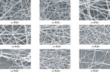
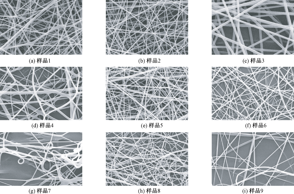
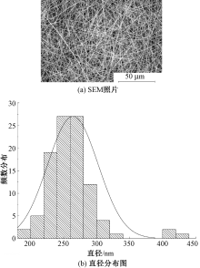
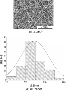
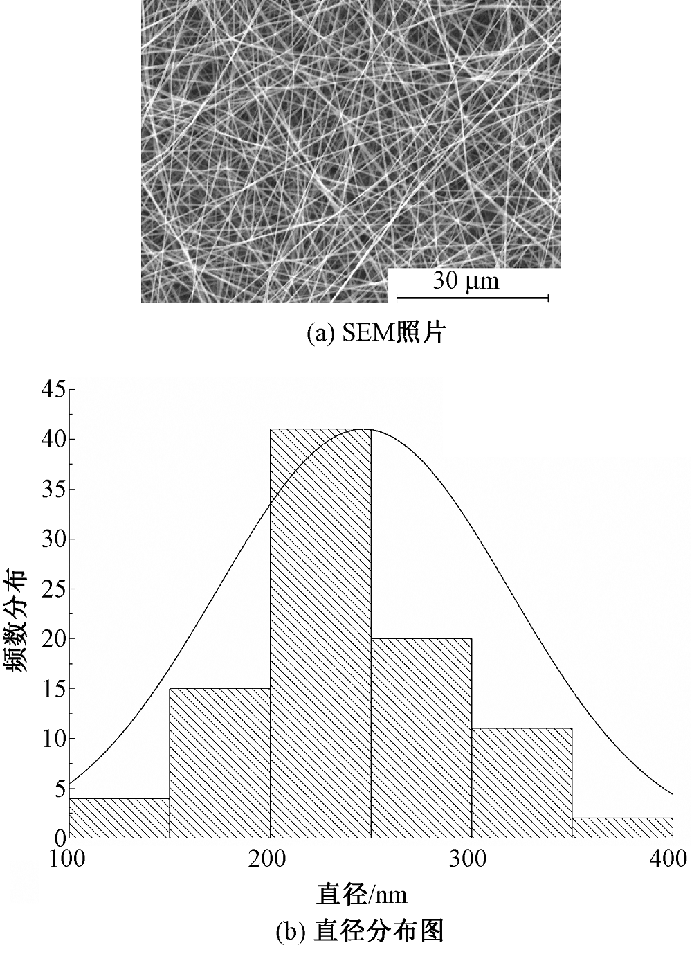
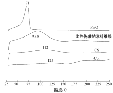
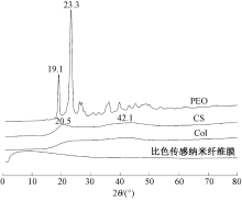
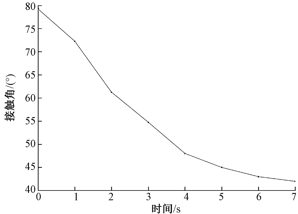
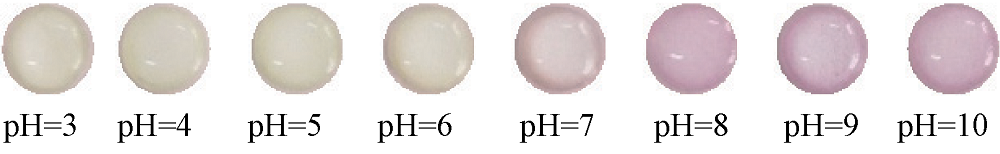
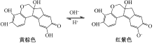
为开发用于监测伤口感染问题的纳米纤维材料,以植物染料苏木为指示剂,壳聚糖、鱼胶蛋白为载体原料,利用静电纺丝技术制备了比色传感纳米纤维膜,并对静电纺丝参数进行优化,借助扫描电子显微镜、差示扫描量热仪和X射线衍射仪对纳米纤维膜的微观形貌进行表征,在不同pH值条件下对其变色情况进行探究。结果表明:壳聚糖与鱼胶蛋白质量比为1:1,纺丝电压为12 kV,推进速度为1.5 mL/h,接收距离为20 cm时,制备的纳米纤维膜质量较好,其纤维平均直径为246.2 nm,直径CV值为29.54%;当pH值从5变至7时,比色传感纳米纤维膜的颜色由黄色变为紫色,其变色域与皮肤发炎时渗出液pH值变化一致,符合伤口监测的要求。

关键词: 伤口监测, 纳米纤维膜, 比色传感器, 静电纺丝, 苏木, 壳聚糖, 鱼胶蛋白

Abstract:

Objective The nanofiber membrane structure with high specific surface area and high porosity can be applied in the detection field in response to external stimuli. Recently, the preparation of colorimetric sensors with nanofiber membrane as a carrier has attracted increasing attention. In order to develop nanofiber materials for monitoring wound infection, a colorimetric sensing nanofiber membrane for wound monitoring was prepared from chitosan/fish gum protein as raw material and plant dye hematoxylin as indicator by electrospinning technology.

Method Colorimetric sensing nanofiber membrane was prepared by electrospinning technology after the spinning solution containing hematoxylin was well mixed by the original solution coloring method with optimized parameters of electrospinning. Scanning electron microscope, differential scanning calorimetry and X-ray diffraction (XRD) were dopted to characterize the microscopic morphology of the nanometer fiber membrane and analysis, and the color change situation under different pH values was also studied. In addition, the hydrophilicity of the nanofiber membrane was proved by the hydrophobic angle test.

Results In the prepared nanofiber membrane without beading, the fibers with an average diameter of 346.1 nm were found thin and straight when the mass ratio of chitosan (CS) to collagen (Col) is 1:1 (Fig.1). When the voltage was set to 12 kV, the propulsion speed was 1.5 mL/L, and the receiving distance was 20 cm, the fibers without adhesion phenomenon showed straight and smooth, in which the average fiber diameter was about 264 nm, and the diameter CV value was 14.51% (Tab.2 and Fig.3). Based on the optimal parameters of electrospinning, the colorimetric sensing nanofiber membrane was prepared with hematoxylin (Fig.4). The thickness of the nanofiber membrane was 0.01 mm, in which the average diameter of the fiber was 246.2 nm. The colorimetric sensing nanofiber membrane had a peak of 93.8 ℃, which is lower than CS and Col (Fig.5). This believed to be precise because the melting temperature of polyethylene oxide (PEO) in the nanofiber membrane is lower than that of CS and Col. Therefore, the introduction of PEO reduced the melting temperature of the nanofiber membrane, indicating that the three materials successfully integrated into the nanofiber membrane. Furthermore, the stucture test results showed that the nanofiber membrane reveals a peak near 10°, meanwhile, the peak strength is lower than that of chitosan and collagen (Fig.6). It may be because CS and Col were successfully mixed, which destroys the helical structure of collagen and affects the crystal structure of nanofiber membrane. The contact angle test results showed that the contact angle of the prepared nanofiber membrane changed from 80° to 46° within 4 s, which showed good hydrophilicity and suitability for medical applications (Fig.7). Most importantly, the color changes of colorimetric sensing nanofiber membranes at different pH values (Fig.8). With the increase of pH value, colorimetric sensing nanofiber membranes exhibited different colors. When pH value increased from 5 to 7, the color change was more obvious, the fiber membrane changed from yellow to purple, the color difference from 0 to 10.59, and the color change of nanofiber membrane could be observed by naked eye, which was in line with the need of wound monitoring.

Conclusion When the mass ratio of the CS to Col of 1:1, the spinning voltage is 12 kV, the pushing speed is 1.5 mL/h, and the receiving distance is 20 cm, the nanofiber membrane obtained possess good hydrophilicity. The average fiber diameter is 246.2 nm, and the diameter CV value is 29.54%. More importantly, the color of the colorimetric sensing nanofiber membrane changes from yellow to purple, when the pH value changes from 5 to 7. Moreover, the color change range of the nanofiber membrane is consistent with the pH value of the exudate when the skin is inflamed, which meets the requirements of wound monitoring.

Key words: wound monitoring, nanofiber membrane, colorimetric sensor, electrospinning, hematoxylin, chitosan, fish collagen

中图分类号: 

  • TS159

表1

正交试验因素水平表"

水平 A
电压/kV
B
接收距离/cm
C
推进速度/
(mL·h-1)
1 12 15 0.5
2 16 20 1.0
3 20 25 1.5

图1

CS和CS/Col纳米纤维膜形貌(×10 000)"

表2

正交试验方案及结果"

样品
编号
因素 平均
直径/nm
直径CV
值/%
A/kV B/cm C/(mL·h-1)
1 12 15 0.5 378.4 41.108
2 12 20 1.0 330.1 35.617
3 12 25 1.5 344.5 14.513
4 16 15 1.0 491.7 48.613
5 16 20 1.5 277.0 19.896
6 16 25 0.5 270.0 26.919
7 20 15 1.5 397.0 32.962
8 20 20 0.5 238.9 32.716
9 20 25 1.0 315.4 41.736
K1 1 053.0 1 267.1 887.3
K2 1 038.7 846.0 1 137.2
K3 951.3 929.9 1 018.5
R 33.9 140.4 83.3

图2

正交试验制备的纳米纤维膜形貌(×10 000)"

图3

最优工艺制备的CS/Col纤维膜形貌及直径分布图"

图4

比色传感纳米纤维膜形貌及直径分布图"

图5

比色传感纳米纤维膜及原料的DSC曲线"

图6

比色传感纳米纤维膜及原料的XRD曲线"

图7

比色传感纳米纤维膜动态水接触角"

表3

不同pH值下纳米纤维膜的颜色特征值"

pH值 L a b H/(°) ΔE
3 81.2 1.3 9.4 38 0.98
4 80.7 1.2 8.4 40 1.12
5 80.3 0.9 9.4 42 0.00
6 80.0 2.1 9.4 40 1.24
7 75.9 8.7 3.2 4 10.89
8 72.0 15.5 -3.7 331 21.30
9 71.2 14.9 -3.9 328 21.35
10 70.9 15.3 -3.8 330 21.68

图8

在不同pH值下比色传感纳米纤维膜的变色情况"

图9

苏木红色素变色机制"

[1] 田玉玲. 基于pH变色指示伤口的载药织物的制备及性能研究[D]. 上海: 东华大学, 2014:8-12.
TIAN Yuling. Preparation and properties of drug-loaded fabric based on pH discoloration indicating wound[D]. Shanghai: Donghua University, 2014:8-12.
[2] GAMERITH C, LUSCHNING D, ORTHER A, et al. pH-responsive materials for optical monitoring of wound status[J]. Sensors and Actuators B:Chemical, 2019. DOI:10.1016/j.snb.2019.126966.
doi: 10.1016/j.snb.2019.126966
[3] 陈莉, 李潇. 姜黄染色织物的pH值敏感变色性能[J]. 纺织学报, 2017, 38(4): 80-84.
CHEN Li, LI Xiao. pH sensitive color change properties of turmeric dyed fabrics[J]. Journal of Textile Research, 2017, 38(4): 80-84.
[4] 李洋, 张元明, 姜伟, 等. 茜草植物染料染色莫代尔纤维的超声波处理[J]. 纺织学报, 2019, 40(4): 83-89.
LI Yang, ZHANG Yuanming, JIANG Wei, et al. Ultrasonic treatment of madder fiber dyed with rubia plant dye[J]. Journal of Textile Research, 2019, 40(4): 83-89.
[5] 卢声. 固色和铁媒染对苏木染色丝织物颜色稳定性的影响[J]. 纺织学报, 2013, 34(10): 96-100.
LU Sheng. Effect of fixing color and iron dyeing on color stability of sappanwood dyed silk fabrics[J]. Journal of Textile Research, 2013, 34(10): 96-100.
[6] XU W, HU X, ZHUANG S, et al. Flexible and salt resistant janus absorbers by electrospinning for stable and efficient solar desalination[J]. Advanced Energy Materials, 2018. DOI:10.1002/aenm.201702884.
doi: 10.1002/aenm.201702884
[7] WEI Zhiqiang, ZHOU Zhankai, LI Qiuyu, et al. Flexible nanowire cluster as a wearable colorimetric humidity sensor[J]. Small, 2017. DOI:10.1002/SMLL.201700109.
doi: 10.1002/SMLL.201700109
[8] 孙倩, 阚燕, 李晓强, 等. 聚丙烯腈/氯化钴纳米纤维比色湿度传感器的制备及其性能[J]. 纺织学报, 2020, 41(11): 27-33.
SUN Qian, KAN Yan, LI Xiaoqiang, et al. Preparation and performance of polyacrylonitrile/cobalt chloride nanofiber colorimetric humidity sensor[J]. Journal of Textile Research, 2020, 41 (11): 27-33.
[9] 倪安琪, 高婧, 侯兵兵, 等. 变色响应聚丙烯腈纳米纤维膜的制备及其过滤性能[J]. 东华大学学报(自然科学版), 2017, 43(6): 771-778.
NI Anqi, GAO Jing, HOU Bingbing, et al. Preparation and filtration performance of color responsive polyacrylonitrile nanofiber membrane[J]. Journal of Donghua University (Natural Science), 2017, 43 (6): 771-778.
[1] 胡蝶飞, 王琰, 姚菊明, 祝国成. 纳米纤维复合结构空气过滤材料性能研究[J]. 纺织学报, 2023, 44(05): 77-83.
[2] 周堂, 汪邓兵, 赵磊, 刘祖一, 凤权. 负载WO3的细菌纤维素/Au膜制备及其催化性能[J]. 纺织学报, 2023, 44(04): 16-23.
[3] 李好义, 贾紫初, 刘宇亮, 谭晶, 丁玉梅, 杨卫民, 牟文英. 高压静电加载形式对聚合物熔体静电直写制备效果的影响[J]. 纺织学报, 2023, 44(04): 32-37.
[4] 张少月, 岳江昱, 杨家乐, 柴晓帅, 冯增国, 张爱英. 环境友好聚己内酯基复合相变纤维膜的制备及其性能[J]. 纺织学报, 2023, 44(03): 11-18.
[5] 陈萌, 何瑞东, 程怡昕, 李纪伟, 宁新, 王娜. 磁控溅射银/锌改性聚苯乙烯/聚偏氟乙烯复合纤维膜的制备及其性能[J]. 纺织学报, 2023, 44(03): 19-27.
[6] 何满堂, 王黎明, 覃小红, 俞建勇. 静电纺纳米纤维在界面太阳能蒸汽转化应用中的研究进展[J]. 纺织学报, 2023, 44(03): 201-209.
[7] 杨广鑫, 张庆乐, 李小超, 李思瑜, 陈辉, 程璐, 夏鑫. 热诱导熔接聚氨酯/聚二甲基硅氧烷防水透湿膜的制备及其性能优化[J]. 纺织学报, 2023, 44(03): 28-35.
[8] 葛铖, 郑元生, 刘凯, 辛斌杰. 电压对静电纺串珠纤维成形过程的影响[J]. 纺织学报, 2023, 44(03): 36-41.
[9] 周泠卉, 曾佩, 鲁瑶, 付少举. 聚乙烯醇纳米纤维膜/罗纹空气层织物复合吸声材料的制备及其性能[J]. 纺织学报, 2023, 44(03): 73-78.
[10] 周歆如, 胡铖烨, 范梦晶, 洪剑寒, 韩潇. 双针头连续水浴静电纺的电场模拟及其纳米纤维包芯纱结构[J]. 纺织学报, 2023, 44(02): 27-33.
[11] 周文, 俞建勇, 张世超, 丁彬. 基于绿色溶剂的聚酰胺纳米纤维膜制备及其空气过滤性能[J]. 纺织学报, 2023, 44(01): 56-63.
[12] 方寅春, 陈吕鑫, 李俊伟. 阻燃超疏水涤/棉混纺织物的制备及其性能[J]. 纺织学报, 2022, 43(11): 113-118.
[13] 张长欢, 李纤纤, 张力冉, 李德阳, 李念武, 吴红艳. 磷酸铁锂/炭黑/碳纳米纤维柔性正极的制备及其性能[J]. 纺织学报, 2022, 43(11): 16-21.
[14] 王洪杰, 胡忠文, 王赫, 凤权, 林童. 单向导湿纺织品及其应用的研究进展[J]. 纺织学报, 2022, 43(11): 195-202.
[15] 吴焕岭, 谢周良, 汪阳, 孙万超, 康正芳, 徐国华. 胶原蛋白改性聚乳酸-羟基乙酸载药纳米纤维膜的制备及其性能[J]. 纺织学报, 2022, 43(11): 9-15.
Viewed
Full text


Abstract

Cited

  Shared   
  Discussed   
No Suggested Reading articles found!