纺织学报 ›› 2024, Vol. 45 ›› Issue (07): 18-23.doi: 10.13475/j.fzxb.20230301001
于承浩1,2,3, 王元非1, 于腾波2, 吴桐1,3( )
)
YU Chenghao1,2,3, WANG Yuanfei1, YU Tengbo2, WU Tong1,3()
摘要:
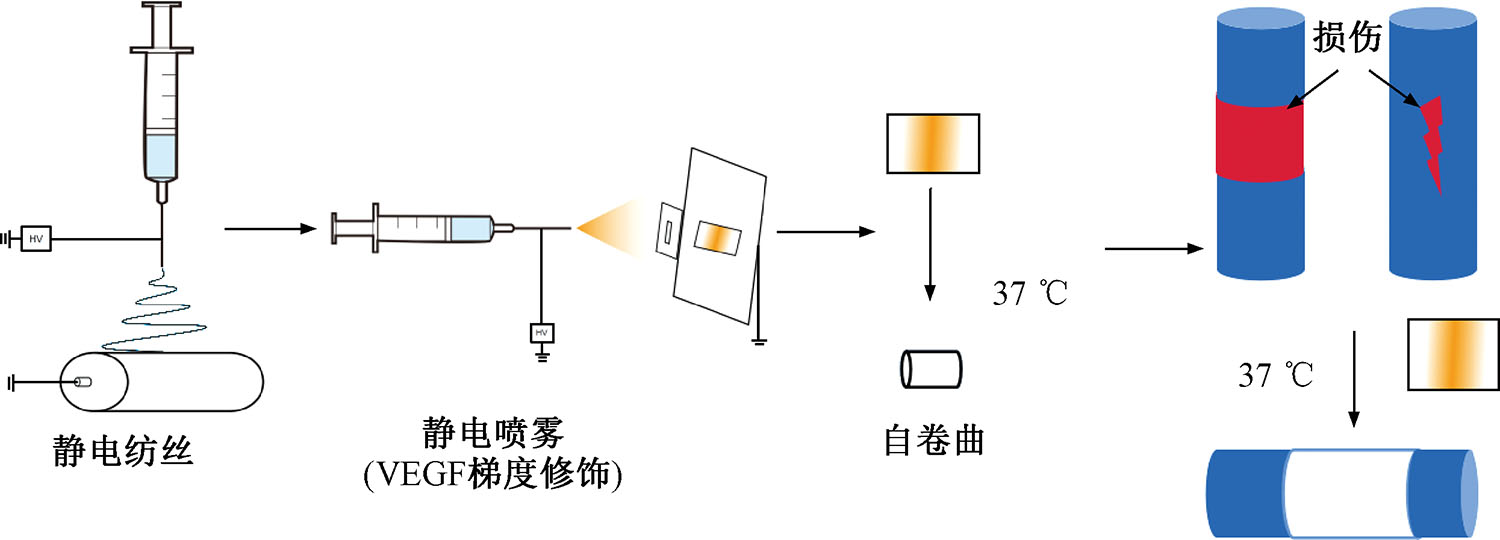
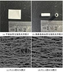
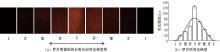
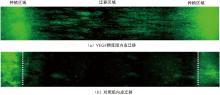
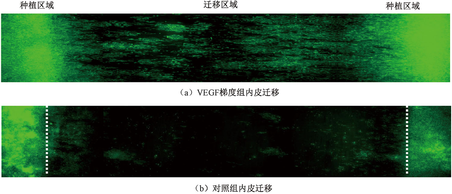
为研究热致自卷曲与生长因子梯度化修饰对血管内皮化的促进作用,使用左旋聚乳酸(PLLA)和聚乳酸-羟基乙酸共聚物(PLGA)作为材料,通过静电纺丝技术制备了具有热致自卷曲特性的PLLA/PLGA纳米纤维血管支架。通过静电喷雾技术制备了梯度生长因子修饰血管支架内层,并对其自卷曲、微观结构、生物相容性及内皮化功能进行表征。结果表明:制备的PLLA/PLGA血管支架具备多层取向纳米纤维结构,厚度为(6.75 ± 0.4) μm,具有出色的生物相容性,可在37 ℃条件下自卷曲成管状结构;血管支架内层膜对生长因子成功进行了梯度修饰,修饰后血管支架的细胞迁移距离是未修饰的3.5倍,从而加快了内皮细胞迁移,促进血管内层的快速内皮化。
中图分类号:
| [1] | GHAVAMI H S, KHOSHTINAT M, SADEGHI-FARAH S, et al. The relationship of coffee consumption and CVD risk factors in elderly patients with T2DM[J]. BMC Cardiovascular Disorders, 2021. DOI:10.1186/s12872-021-02058-7. |
| [2] | GOMES C P C, SCHROEN B, KUSTER G M, et al. Regulatory RNAs in heart failure[J]. Circulation, 2020,141:313-328. |
| [3] | GONCALVES R C, BANFI A, OLIVEIRA M B, et al. Strategies for re-vascularization and promotion of angiogenesis in trauma and disease[J]. Biomaterials, 2021. DOI: 10.1016/j.biomaterials.2020.120628. |
| [4] | JIN D, HU J, XIA D, et al. Evaluation of a simple off-the-shelf bi-layered vascular scaffold based on poly(L-lactide-co-ε-caprolactone)/silk fibroin in vitro and in vivo[J]. International Journal of Nanomedicine, 2019,14:4261-4276. |
| [5] | AWAD N K, NIU H, ALI U, et al. Electrospun fibrous scaffolds for small-diameter blood vessels: a review[J]. Membranes, 2018. DOI: 10.3390/membranes8010015. |
| [6] | LIU R H, ONG C S, FUKUNISHI T, et al. Review of vascular graft studies in large animal models[J]. Tissue Engineering Part B: Reviews, 2018,24:133-143. |
| [7] | 孙子茹, 焦恩祥, 袁坤山, 等. 静电纺丝在构建小口径仿生人工血管中的研究进展[J]. 材料导报, 2023, 37(S1):486-490. |
| SUN Ziru, JIAO Enxiang, YUAN Kunshan, et al. Research progress of electrostatic spinning in the construction of bionic small-diameter artificial blood vessels[J]. Materials Review, 2023, 37(S1):486-490. | |
| [8] | BUSCEMI S, PALUMBO V D, MAFFONGELLI A, et al. Electrospun PHEA-PLA/PCL scaffold for vascular regeneration: a preliminary in vivo evaluation[J]. Transplantation Proceedings, 2017,49:716-721. |
| [9] | FARTO-VAAMONDE X, XIAZ-GOMEZ L, PARGA A, et al. Perimeter and carvacrol-loading regulate angiogenesis and biofilm growth in 3D printed PLA scaffolds[J]. J Control Release, 2022,352:776-792. |
| [10] | BAZGIR M, SAEINASAB M, ZHANG W, et al. Investigation of cell adhesion and cell viability of the endothelial and fibroblast cells on electrospun PCL, PLGA and coaxial scaffolds for production of tissue engineered blood vessel[J]. J Funct Biomater, 2022. DOI: 10.3390/jfb13040282. |
| [11] | HU X, SHEN H, YANG F, et al. Preparation and cell affinity of microtubular orientation-structured PLGA(70/30) blood vessel scaffold[J]. Biomaterials, 2008,29:3128-3136. |
| [12] | 刘月, 蒋紫仪, 李晶晶, 等. 聚左旋乳酸己内酯/丝素蛋白小口径人工血管细胞共培养及体内生物相容性[J]. 中国组织工程研究, 2022, 26(22):3505-3513. |
| LIU Yue, JIANG Ziyi, LI Jingjing, et al. Cell co-culture and in vivo biocompatibility of poly(l-lactic caprolactone)/silk fibroin small-diameter artificial blood vessels[J]. Chinese Journal of Tissue Engineering Research, 2022, 26(22):3505-3513. | |
| [13] | 吴桐, 王元非, 于承浩, 等. 一种热致自卷曲聚合物管状支架及其制备方法: 202310102575.3[P].2023-04-25. |
| WU Tong, WANG Yuanfei, YU Chenghao, et al.A thermally induced self curling polymer tubular scaffold and its preparation method: 202310102575.3[P].2023-04-25. | |
| [14] | HUANG W, TONG Z, WANG R, et al. A review on electrospinning nanofibers in the field of microwave absorption[J]. Ceramics International, 2020, 46(17):26441-26453. |
| [15] | CHOR A, GONCALVES R P, COSTA A M, et al. In vitro degradation of electrospun poly(lactic-co-glycolic acid) (PLGA) for oral mucosa regeneration[J]. Polymers (Basel), 2020. DOI: 10.3390/polym12081853. |
| [16] | ZHANG X, XU X L, CHEN L S, et al. Multi-responsive hydrogel actuator with photo-switchable color changing behaviors[J]. Dyes and Pigments, 2020. DOI:10.1016/j.dyepig. 2019.108042. |
| [17] | 郭凤云, 过子怡, 高蕾, 等. 热粘结复合纤维人造血管支架的制备及其性能[J]. 纺织学报, 2021, 42(6): 46-50. |
| GUO Fengyun, GUO Ziyi, GAO Lei, et al. Preparation and properties of thermal bonded fibrous artificial blood vessels[J]. Journal of Textile Research, 2021, 42(6):46-50. | |
| [18] | BEHESHTIZADEH N, AZAMI M, ABBASI H, et al. Applying extrusion-based 3D printing technique accelerates fabricating complex biphasic calcium phosphate-based scaffolds for bone tissue regenera-tion[J]. Journal of Advanced Research, 2022,40:69-94. |
| [19] | HAN F, JIA X, DAI D, et al. Performance of a multilayered small-diameter vascular scaffold dual-loaded with VEGF and PDGF[J]. Biomaterials, 2013,34:7302-7313. |
| [20] | FIORETTA E S, FLEDDERUS J O, BURAKOWSKA-MEISE E A, et al. Polymer-based scaffold designs for in situ vascular tissue engineering: controlling recruitment and differentiation behavior of endothelial colony forming cells[J]. Macromolecular Bioscience, 2012,12:577-590. |
| [21] | APTE R S, CHEN D S, FERRARA N. VEGF in signaling and disease: beyond discovery and development[J]. Cell, 2019, 17:1248-1264. |
| [22] | WU T, ZHANG J, WANG Y, et al. Fabrication and preliminary study of a biomimetic tri-layer tubular graft based on fibers and fiber yarns for vascular tissue engineering[J]. Mater Sci Eng C:Mater Biol Appl, 2018,82:121-129. |
| [23] | PERTICI V, MARTRIU G, GIMES D, et al. Synthetic polymer-based electrospun fibers: biofunctionalization strategies and recent advances in tissue engineering, drug delivery and diagnostics[J]. Current Medicinal Chemistry, 2018,25:2385-2400. |
| [24] | SPADACCIO C, NAPPI F, DE MARCO F, et al. Preliminary in vivo evaluation of a hybrid armored vascular graft combining electrospinning and additive manufacturing techniques[J]. Drug Target Insights, 2016,1:1-7. |
| [1] | 于雯, 邓南平, 唐湘泉, 康卫民, 程博闻. 静电溶吹微纳无机纤维制备技术及其应用进展[J]. 纺织学报, 2024, 45(07): 230-239. |
| [2] | 刘思彤, 金丹, 孙东明, 李懿轩, 王艳慧, 王静, 王原. 静电纺纳米纤维结构的研究进展[J]. 纺织学报, 2024, 45(06): 201-209. |
| [3] | 徐振凯, 马鸣, 蔺多佳, 刘航, 张剑峰, 夏鑫. 自支撑聚吡咙基碳纤维负极材料的制备及其电化学性能[J]. 纺织学报, 2024, 45(06): 23-31. |
| [4] | 时吉磊, 陈廷彬, 付少海, 张丽平. 低红外发射率控温热红外伪装材料的制备与性能[J]. 纺织学报, 2024, 45(06): 32-38. |
| [5] | 栗志坤, 于影, 左雨欣, 史豪秦, 金玉珍, 陈洪立. 聚丙烯腈/二硫化钼复合薄膜的挠曲电效应分析及其应用[J]. 纺织学报, 2024, 45(05): 27-34. |
| [6] | 梁文静, 吴俊贤, 何崟, 刘皓. 基于复合纳米纤维膜的离子传感器制备及其性能[J]. 纺织学报, 2024, 45(04): 15-23. |
| [7] | 宋贝贝, 赵浩阅, 李欣宇, 屈展, 方剑. 载有MXene的钴氮掺杂碳纳米纤维在锂硫电池中的应用[J]. 纺织学报, 2024, 45(04): 24-32. |
| [8] | 贾琳, 董晓, 王西贤, 张海霞, 覃小红. 聚己内酯/MgO复合纳米纤维膜的制备及其性能[J]. 纺织学报, 2024, 45(04): 59-66. |
| [9] | 陆瑶瑶, 叶俊涛, 阮承祥, 娄瑾. 二氧化钛/多孔碳纳米纤维复合材料的制备及其光催化性能[J]. 纺织学报, 2024, 45(04): 67-75. |
| [10] | 杨琪, 邓南平, 程博闻, 康卫民. 树枝状磺化聚醚砜纤维基复合固态电解质的制备及其性能[J]. 纺织学报, 2024, 45(03): 1-10. |
| [11] | 赵美奇, 陈莉, 钱现, 李晓娜, 杜迅. 用于铜离子检测的静电纺纤维膜制备及其性能[J]. 纺织学报, 2024, 45(03): 11-18. |
| [12] | 丁彩红, 顾馨, 卢晨雨. 血管支架预制件的六角形三维虚拟编织[J]. 纺织学报, 2024, 45(03): 65-73. |
| [13] | 田博阳, 王向泽, 杨湙雯, 吴晶. 非对称结构纤维膜的制备及其热调控性能[J]. 纺织学报, 2024, 45(02): 11-20. |
| [14] | 周歆如, 范梦晶, 岳欣琰, 洪剑寒, 韩潇. 导电微纳纤维复合纱的制备及其气敏特性[J]. 纺织学报, 2024, 45(02): 52-58. |
| [15] | 戎成宝, 孙辉, 于斌. 银-铜双金属纳米粒子/聚乳酸复合纳米纤维膜的制备及其抗菌性能[J]. 纺织学报, 2024, 45(01): 48-55. |
|
||
 京公网安备11010502044800号